Playlist
Show Playlist
Hide Playlist
Thyroid Gland

-
Slides 05 Human Organ Systems Meyer.pdf
-
Download Lecture Overview
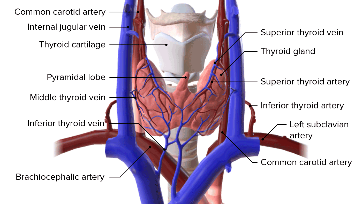
00:00 First, I will describe the thyroid gland. It’s a bilobed gland. It’s in the neck region. You can see labelled here at the top of the left-hand section through the neck region, a lobe of the thyroid. 00:18 It’s separated by the trachea, so one lobe is on either side, joined by a very thin isthmus of thyroid secretory units that I won’t describe in this particular lecture. Trachea is a roughly circular structure. If you look carefully, it has got the cartilage rings that support it. And then the other structure on the far left of this particular section is the esophagus. On the right-hand side, you can see details of the thyroid gland at a higher magnification. If you look very carefully at this particular section on the right-hand side, you can see that it’s made up of structures or functional units of the thyroid we call thyroid follicles. And look very carefully into this section. These thyroid follicles are of very different size. Their diameters range from about 0.2 millimeters to about 1 millimeter. 01:22 And this probably reflects their real size, not necessarily the different state of activity of each follicle. We’ll see this again when we look at the endocrine component of the pancreas. When you look at the follicle, the thyroid follicle under higher magnification, you can see on the left-hand picture more details of the structure of this follicle. 01:51 It has cells in it called follicular cells. These follicular cells are epithelial cells, they're endocrine cells. They form the lining or the wall of each follicle. And within the follicle itself, you can see colloid. This is a gel-like component. It’s actually stored product that consists of thyroglobulin. These follicular cells secrete that thyroglobulin and store it in the lumen of each follicle. This is an example of the only cell in the body, the only endocrine cell that actually synthesizes its hormonal product and stores it extracellularly outside the cell. These are cuboidal type epithelial cells. But have a look at the very edge of the cells or inside some of the cells you see, and you can see some little tiny dot specs. The edge of the cell is seem to be a little bit sort of unusual or uneven. This represents structures that I’ll show you and talk about in a later slide. This slide shows a diagram of two cells, two follicular cells in the thyroid follicle. 03:14 The green colored component of each of the cells in the diagram at the top represents the colloid. You saw it stained pink, actually, in the previous slides. Remember that colloid stores thyroglobulin. When that thyroglobulin is synthesized on the rough or granular endoplasmic reticulum inside the cell, and then it’s glycosylated as it passes through the endoplasmic reticulum, and also the Golgi apparatus shown in blue. That Golgi apparatus then packages the thyroglobulin into vesicles, which move to the surface of the cell and are released into the space of lumen as colloid by a process called exocytosis. These follicular cells also have microvilli. So that surface that I explained to you earlier in the slide, on the light microscope picture when I said they had little specs, little uneven boundaries close to the lumen, that represents all these exocytotic activity and the little vesicle stored at the apex of the cell on their way to being deposited into the colloid. 04:38 On the right-hand side, you can see that the cell is representing the absorption, the endocytosis of the colloid. The microvillus projections wrap around segments of the colloid. And then by endocytosis, that colloid is then brought inside the cell, where it fuses with lysosomes again at the apex of the cell. And again, this sort of activity is reflected in the uneven surface that you see when you look at the surface of the cells I pointed out in the light microscope pictures. That endocytosed thyroglobulin then reacts with the lysosomal enzymes and other enzymes in the cell, and then the thyroid hormones, T3, and T4, are released on the basal aspect of the cell away from the colloid or the luminal surface into blood capillaries that passed by. In this slide, again, it brings the two previous groups of slides together. On the left-hand side, you see the light microscope picture taken through thyroid follicles. You see the follicular cells labeled and the colloid thyroglobulin. 06:07 And look carefully again at the surface of these cells, and you can often see little specs or unevenness that reflect what you see in the diagram on the right-hand side. In the middle slide, in the middle diagram, I explained how the thyroglobulin is produced and deposited in the colloid, and then how it’s reabsorbed by endocytosis from that colloid into the cell to then be broken down and transformed into the two thyroid hormones, T3, and T4. 06:43 Another step in the process that I want to explain at this point is that iodide is absorbed into the cell and highly concentrated. And that iodide is then oxidized and passed into the colloid as iodine. And that forms a complex with the thyroglobulin, a very important complex which when it’s reabsorbed back into this cell, as I’ve explained before, contributes to the formation of the thyroid hormones. So two very important functions of this cell, one, to produce the thyroglobulin, and one to concentrate and oxidize iodide that helps form this iodinated thyroglobulin complex inside the colloid. Another very important feature of this slide is if you again look at the light microscope picture, you see a darker pink component running really between the walls of two thyroid follicles. That dark pink component happens to be red blood cells, moving through in capillaries. And as I mentioned before, the thyroid follicular cells, they produce the hormone product and secrete them basally, on the basal aspect of their cells into these capillaries. And it’s important to point out here, and you certainly cannot see it, but these capillaries are very fenestrated. 08:19 And this is a typical type of capillary you see in endocrine glands. Fenestrations allow very easy movement of fluid from the interstitium which contains initially the hormonal product. 08:32 It enables that fluid to pass in to the blood, and therefore, be carried to the rest of the body. There is another type of cell in a thyroid follicle, squeezed inside the epithelium within the basal lamina or basement membrane around the follicular epithelium. And this is called the C-cell or the parafollicular cells. They have a rather foamy vesicle dominated cytoplasm. 09:05 They secrete calcitonin which is responsible for lowering calcium levels in the blood. By means I will describe in another lecture on the bone, on bone growth and remodeling.
About the Lecture
The lecture Thyroid Gland by Geoffrey Meyer, PhD is from the course Endocrine Histology.
Included Quiz Questions
The parafollicular cells in the thyroid secrete which of the following hormones?
- Calcitonin
- Thyroid hormone
- Insulin
- Somatostatin
- Cortisol
Which of the following is the main function of calcitonin?
- Reducing blood levels of calcium
- Increasing blood level of phosphate
- Increasing urine calcium
- Reducing urine calcium
- Regulating vitamin D absorption
Thyroid follicles are most commonly lined by which of the following types of epithelial cells?
- Cuboidal
- Squamous
- Columnar
- Pseudostratified squamous
- Pseudostratified columnar
Where in the thyroid cell is thyroglobulin initially synthesized?
- Endoplasmic reticulum
- Lysosomes
- Nucleus
- Golgi apparatus
- Peroxisome
The thyroid gland mainly produces what type of hormones?
- Endocrine
- Paracrine
- Autocrine
- Exocrine
- Steroid
Author of lecture Thyroid Gland

Geoffrey Meyer, PhD
Customer reviews
5,0 of 5 stars
| 5 Stars |
|
1 |
| 4 Stars |
|
0 |
| 3 Stars |
|
0 |
| 2 Stars |
|
0 |
| 1 Star |
|
0 |
liked it because it was easy to follow. professor gives you a chance to absorb information.
