Playlist
Show Playlist
Hide Playlist
Bowel Obstruction and Ileus: Ileus & Small Bowel Obstruction

-
Slides Ileus and Obstruction.pdf
-
Download Lecture Overview
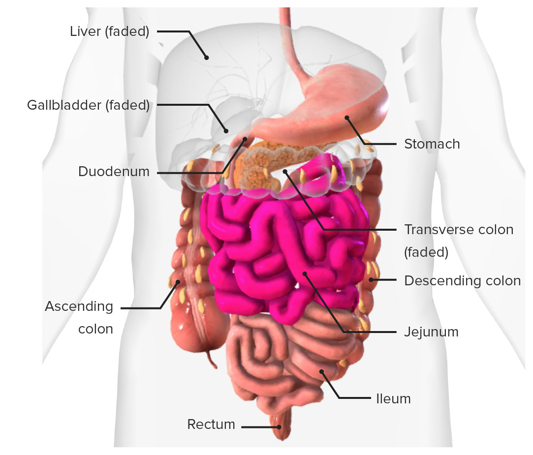
00:01 So one of the most common reasons for performing an abdominal imaging study is to evaluate for bowel obstruction or ileus. 00:07 So let's take a look at these findings that we would see. 00:10 This is 25-year-old patient that came in with the right lower abdominal pain. 00:14 It's a female patient. 00:16 Let's take a look at this case and see which you can see here and keep this in mind until we get through the lecture and then we'll go back to this at the very end. 00:24 So what do you look for when you're looking for a bowel obstruction? You wanna look for dilated loops of large or small bowel. 00:33 You wanna see if it's focal or diffuse. 00:36 You wanna take a look at air fluid levels. 00:38 You wanna see if there's air within the rectum, and you wanna see if there's a transition point when you're performing a CT. 00:46 So what's the difference between a functional ileus and the mechanical obstruction. 00:50 A functional ileus is loss of normal peristaltic function due to irritation. 00:55 A mechanical obstruction is a physical obstruction in the small or large bowel and this maybe partial or it maybe complete. 01:02 A functional ileus maybe localized or generalized and a mechanical obstruction as we said could be either complete or partial. 01:10 A functional ileus usually affects the small bowel and it doesn't tend to have a transition point while with the mechanical obstruction you do have a transition point and this is the point that which the bowel changes caliber from being dilated to being collapsed. 01:24 The small bowel caliber is typically less than 3cm on a functional ileus. 01:28 So it's not significantly dilated. 01:31 In a mechanical obstruction, the small bowel can become significantly dilated and the caliber usually more than about 3cm. 01:38 In a functional ileus you would see air within the rectum because the air continues to pass past the site of aperistalsis while in a mechanical obstruction if you have a complete obstruction you would have no air within the rectum because nothing is getting pass that point of obstruction. 01:54 So a partial or early small bowel obstruction within the first day or two can appear identical to an ileus. So it's actually very hard to differentiate and usually what we do is follow up imaging to see how it evolves. 02:06 So in a localized functional ileus, you would have dilatation of a loop or few loops of small bowel and these are called the sentinal loops. 02:17 This is cause by adjacent irritation. 02:19 It can result from really any kind of abdominal pathology that causes focal inflammation. 02:24 Types of pathologies include cholecystities, pancreatitis, appendicitis, diverticulitis or even ureteral stones. 02:35 So anything that causes pain in the patient can cause a localize functional ileus. 02:40 A generalize functional ileus is most often postoperative and it can involve the entire bowel both small and large. 02:48 So if you take a look at this patient here, you have multiple dilated loops of both large and small bowel. 02:53 They're actually so dilated that we really aren't able to tell the difference between large and small bowel and you can see that this patient is postoperative. 03:00 They have surgical staples among the midline of the abdomen. 03:04 A small bowel obstruction is usually caused by a lesion that obstructs the lumen of the bowel. 03:11 It results in dilatation of the bowel proximal to the obstructing lesion and then compression of the bowel after the obstructing lesion. 03:18 This is an example of a normal large bowel and the normal small bowel. 03:25 On the right we have an image of a small bowel obstruction. 03:28 So that large bowel is slightly decrease in caliber because there is less air getting through the point of obstruction which is right here at the terminal ileum. 03:37 The small bowel that's proximal to this part, point of obstruction is dilated. 03:41 There are many different causes of small bowel obstruction. 03:47 The most common include adhesions from prior surgery, tumors, a hernia and we'll take a look at an example of this. 03:55 Inflammatory bowel disease can cause multiple small bowel obstructions. 03:59 Intussusceptions which is bowel tunneling within other bowel. 04:03 It's kinda like of telescoping effect where a piece of bowel tunnels within another piece of bowel and can cause an obstruction at that level. 04:10 Intraluminal lesions other than tumors can also cause small bowel obstruction and they can be from a variety of different reasons including an ingested foreign body, you can have a gallstones ileus which when a gallstone penetrates through the gallbladder wall and enters the small bowel and causes an obstruction within the small bowel or you can have bezoars which are abnormal collection that the patient maybe ingests, certain psychiatric patients may ingest different types of foreign bodies that can collect together and cause an obstruction. 04:38 So this is an example of a patient that's presenting with abdominal pain. 04:44 Let's take a look at this film. 04:47 So how does this bowel gas pattern look? This patient has a prominent loop of small bowel in the left upper abdomen. 05:03 The differential of this includes a focal ileus or an early or partial small bowel obstruction. 05:09 As we said it's really hard to differentiate in a patient that's presenting with symptoms that lasted less than one to two days because early on both can have similar imaging findings. 05:18 The following day however the patient had a repeat film because they had increasing abdominal pain. 05:27 So what do you see on these films here? Here we have a supine film and in this one is an upright film. 05:35 So as you can see there's increasing dilatation of multiple loops of small bowel. 05:42 Now, a majority of the small bowel loops appear dilated, you can see that on the supine film. 05:47 You also have increasing air fluid levels which we can see best on this upright film. 05:53 This is very concerning for a small bowel obstruction. 05:57 So what else can you perform to help you figure out why this is occurring? So in this patient the CT scan was performed, here we have two axial images from abdominal CT scan and we have a coronal image. So let's take a look at each of these images. 06:14 Here, on this axial CT image we have, what looks like a little outpouching of the anterior abdominal wall that contains a loop of bowel. 06:24 On this next image, we again have a loop of bowel that looks a little bit dilated in the same region as the outpouching and it has a modeled appearance and here we have a decompressed loop of bowel. 06:36 On this coronal image you can see a large loop of small bowel that appears dilated. 06:43 So this is a thickened loop of bowel within a ventral hernia that's causing a bowel obstruction and here we have a transition point at the level of obstruction. This modeled loop of small bowel here is what we call the small bowel feces sign. 07:01 So you normally should not have any stool within the small bowel when you do have stool within the small bowel you should suspect a small bowel obstruction and that's called the small bowel feces sign. 07:11 So what are some CT findings of small bowel obstruction? You should see a dilated loops of small bowel, greater than about 3cm. 07:20 You may or may not see a transition point, you should see collapsed bowel distal to the transition point and you may see the small bowel feces sign. 07:30 So what is a closed loop obstruction? This is an obstruction of a loop of bowel in two separate places. 07:38 You normally don't see any significant dilatation proximal to the closed loop and often this is caused by adhesions from prior surgery. 07:47 This can actually result in strangulations, so this is a surgical emergency, it can cause blocked blood flow to the bowel which can result in necrosis. 07:56 On imaging we see a thickened wall and you may see decreased enhancement of the bowel wall. 08:01 So closed loop obstruction needs immediate surgical treatment because of the very high risk of perforation from the strangulation.
About the Lecture
The lecture Bowel Obstruction and Ileus: Ileus & Small Bowel Obstruction by Hetal Verma, MD is from the course Abdominal Radiology. It contains the following chapters:
- Bowel Obstruction and Ileus
- Small Bowel Obstruction
Included Quiz Questions
Which of the following imaging findings are more commonly associated with small bowel obstruction than ileus?
- Lack of air in the rectum
- Small bowel caliber of < 3 cm
- Lack of transition point
- Focal bowel dilatation
- Normal caliber large bowel
Which part of the bowel appears dilated in a distal jejunal mechanical obstruction?
- Proximal jejunum
- Distal jejunum
- Ileum
- Ascending colon
- Descending colon
In an abdominal imaging study, sentinel loops of the small bowel signify...?
- ...localized functional ileus due to adjacent irritation.
- ...generalized functional ileus.
- ...small bowel obstruction by tumor distal to the dilated loops.
- ...intussusception.
- ...hernia.
Which of the following is TRUE regarding small bowel obstruction?
- There may or may not be a transition point.
- Dilated loops of the small bowel can be > 7 cm.
- Feces sign is seen in the colon. .
- In a closed-loop obstruction, there is a significant dilatation proximal to the closed loop.
- A closed-loop obstruction on imaging demonstrates increased enhancement of the bowel wall.
Author of lecture Bowel Obstruction and Ileus: Ileus & Small Bowel Obstruction

Hetal Verma, MD
Customer reviews
5,0 of 5 stars
| 5 Stars |
|
5 |
| 4 Stars |
|
0 |
| 3 Stars |
|
0 |
| 2 Stars |
|
0 |
| 1 Star |
|
0 |
