Playlist
Show Playlist
Hide Playlist
Cell Structure

-
Basic Histology 01.pdf
-
Download Lecture Overview
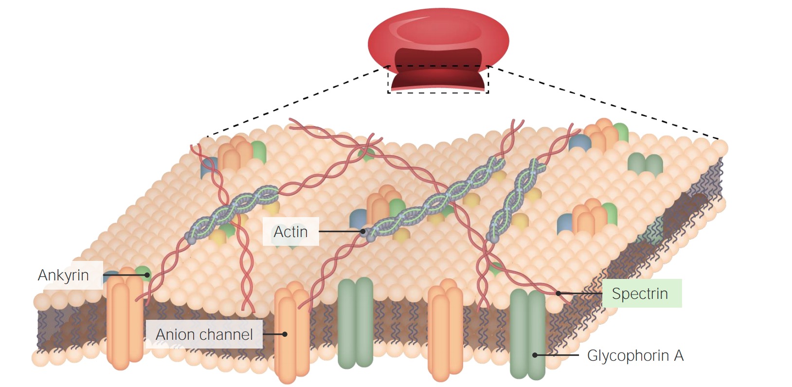
00:01 Let's just define a few terms when we look at the cell. 00:05 Here is a diagram on the right-hand side typical of a mammalian cell or a eukaryote. 00:13 First of all, the term protoplasm. This refers to the nucleus and the cytoplasm, both labeled 1 on the image you see in front of you. Then you have on the outside, the cell membrane, labeled 2.The cell membrane is a very, very important structure. 00:35 It is the barrier between the very stable internal environment of the cell and the external environment. 00:46 It allows transfer of materials across the cell membrane. Oxygen, nutrients, waste products, etc. 00:55 can pass in and out of the cell through the cell membrane through a very detailed set of transport processes, and this system whereby there is transfer across the cell wall in the mammalian cell is referred to in Physiology as an open system. 01:15 The cell has a nucleus, and that nucleus has a nuclear envelope around it or a nuclear membrane. 01:23 Within the nucleus is the nucleoplasm that is a series of very fine scaffolding fibers as well as some fluid. 01:35 It contains also chromatin, which really are just strands of DNA, and rather obvious in this image is that semi-spherical structure you see colored in blue. 01:49 That is the nucleolus. It's the typical structure found in mostly all nuclei in the mammalian cell population. 02:01 The cytoplasm consists of the ectoplasm and endoplasm. 02:07 Ectoplasm is just that very fine piece of cytoplasm immediately underneath the cell membrane. 02:14 It doesn't contain any organelles. All it does is contain very fine actin filaments, and those actin filaments help to support the very delicate cell membrane. 02:29 The endoplasm is that part of the cytoplasm that contains all the organelles, those structures I mentioned earlier that are membrane-bound and that serve specific functions in the cell. 02:47 The endoplasm also includes what we call cytoplasmic inclusions. 02:53 Cytoplasmic inclusions are structures that may not be in all cells. They're inclusions that might be peculiar to individual cell populations, and we'll see those when I describe these in more detail in a later lecture. 03:10 And of course, there is the cytosol or the cell sap, cell fluid. And lastly, I just want to briefly mention the cytoskeleton, again, consisting of microtubules, intermediate filaments, and microfilaments and again, let me just repeat that the cytoskeleton is involved with stabilizing the position of organelles, supporting those organelles, even moving them around where necessary, and also supporting the cell membrane. 03:45 Now, this is a picture of your skin. This is the first time you've seen a histological section. 03:52 It shows you the detail of what your skin looks like under a microscope. 03:58 on the bottom left part of the slide, you can see some very bright pink flaky material. That's keratin. 04:07 If you run your finger across the back of your hand like I am here, you can scrape off that keratin. 04:15 That keratin is really the dead products of cells that precede keratinization that form the keratin in the surface layer of cells, and that keratin is lost continuously in our lives. 04:29 That keratin helps to waterproof our body. Underneath that keratin, you can see layers of cells. 04:37 Mostly, you just see circular pale-staining nuclei of the cells, and then further into the slide towards the right-hand side, a much lighter region, that's the dermis of your skin. 04:51 That is supporting connective tissue. 04:54 Without going through all the details—and we don't want to do that in this initial lecture. You don't need to know the details of all this skin here—but what I want you to understand is you can see detail in this slide. 05:10 You can see details of the histology of what your skin looks like. You can see that detail because you use a tool in histology called a microscope, and a microscope does two things. 05:27 It magnifies or zooms in a slide of your tissue such as skin and it also provides you detail of what's in that slide. 05:40 And first of all, you need to cut a very thin section of your skin to be able to magnify it and also to see the detail. Here is a nice picture of the earth and the moon, and you can see in that picture some of the continents. 06:03 Lights are on in many of the cities in those continents. You can't see the details of those lights. 06:12 You can't even see sometimes the difference between lights in one city and another. 06:18 And if I blow up or magnify that picture, you're still not going to see any more detail. 06:23 Magnification doesn't provide any more detail of what you see unless your optical system also improves what we call resolution or what we call finding the detail. 06:39 If we zoom in on our world and get closer, we can see more detail. 06:45 We start to see buildings and cities. We start to see in this lovely city where I'm giving these lectures in histology, Leipzig in Germany, you can start to see the streets and cars. 07:02 And if we zoom in more, if we increase the magnification and have the oculus system to see detail, you start to see writing, the clock on the Town Hall, again, in this lovely city of Leipzig. 07:20 So what I really mean is for you to look at all the histological sections you're going to look at in your histology course, you need to use a microscope that is going to magnify what is in the section and it's going to provide detail. It has the resolving power to distinguish objects that are very, very close together and our eyes can only distinguish objects that are about 0.2 mm apart. 07:54 If I look at my skin, I can't see any objects that are closer than 0.2 mm or 200 µm apart. 08:07 The light microscope enables you to look at detail as you see in this section and you can see objects that are as close as 0.2 µm apart. Again, that's what I mean by greater resolving power using a microscope.
About the Lecture
The lecture Cell Structure by Geoffrey Meyer, PhD is from the course The Mammalian Cell.
Included Quiz Questions
Of the following choices, what is the smallest scale observable with a light microscope?
- 2 micrometers
- 2.0 nanometers
- 0.2 nanometers
- 30 micrometers
The smallest scale observable by the human eye with 20/20 vision is best indicated by which of the following values?
- 0.2 millimeters
- 2.0 nanometers
- 0.2 nanometers
- 0.2 micrometers
- 3 micrometers
Author of lecture Cell Structure

Geoffrey Meyer, PhD
Customer reviews
5,0 of 5 stars
| 5 Stars |
|
2 |
| 4 Stars |
|
0 |
| 3 Stars |
|
0 |
| 2 Stars |
|
0 |
| 1 Star |
|
0 |
Es una excelente conferencia parte desde lo básico para entender la histología que es la biología lo recomiendo para estudiantes que están haciendo la materia de histología y biología del primer año de la carrera de medicina .Muchas gracias por esta gran conferencia
well put together esp the simple intro description of the skin
