Playlist
Show Playlist
Hide Playlist
Tricuspid Atresia


-
Slides Cyanotic Heart Disease.pdf
-
Reference List Pediatric Nursing.pdf
-
Download Lecture Overview
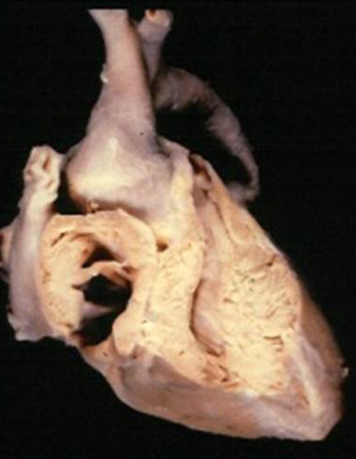
00:01 Let's move on to tricuspid atresia. 00:06 Tricuspid atresia - here, you can see an example on the left part of your slide, you can see a normal heart and on the right side you can see tricuspid atresia. 00:16 These patients are typically very sick when they're born. 00:20 As you can see in a patient with tricuspid atresia, the blood comes back from the IVC and the SVC into the right atrium, and that blood cannot get into that right ventricle. 00:33 It just can't get in - that valve is atretic. 00:37 As a result, that increased pressure causes the blood to blow over the patent foramen ovale into the left side. 00:44 Now, that patient's blood will be able to get out to the body creating a cyanotic infant. 00:52 Some of that blood will get back across into the right venticle and get to the pulmonary artery typically through a VSD. 01:00 These patients, like I said, are very sick and will require an operation right about the time of birth. 01:07 Typically, these patients - much like hypoplastic left heart syndrome, which is another disease we'll talk about in the obstructive cardiac lesions talk - these infants require a gradual switch into single ventricle physiology. 01:24 These infants will be transitioned into only having one ventricle - in this case, it would be the left ventricle - powering the blood to the body. 01:33 Eventually, by the time the arterial switch operation is completed, they'll have passive blood flow that does not go through the heart at all that returns all blood from their body to their lungs. 01:45 This is a 3-stage operation. 01:48 Typically at birth, they have something called a Blalock-Taussig shunt placed. 01:53 The Blalock-Taussig shunt is sort of like a continuing patent ductus arteriosus between the pulmonary artery and the aorta, which allows that mixing to continue to happen. 02:07 Then around a year of life, they'll have a hemi-Fontan, which is another surgical procedure where some of the blood will drain into the pulmonary artery directly - usually from the SVC. 02:21 And then maybe a few years of life, they'll complete the operation and have the inferior vena cava also going into the pulmonary artery. 02:30 It's a gradual stage repair that ends up with an infant with a single ventricle powering the blood to their body and passive blood pressure filling the lungs. 02:41 So remember, with tricuspid atresia, there's no connection between the right atrium and the right ventricle. 02:48 The right ventricle is usually very small and not very effective. 02:53 A VSD is present because, otherwise, the blood can't get back up into the lungs, unless there's a patent ductus arteriosus, which there usually is.
About the Lecture
The lecture Tricuspid Atresia by Brian Alverson, MD is from the course Pediatric Cardiology.
Included Quiz Questions
Which of the following defects is present in tricuspid atresia?
- A small right ventricle
- Aortic stenosis
- Coarctation of the aorta
- A deletion of the p arm of the 5th chromosome
- A closed foramen ovale
Of the three surgeries required for a patient with tricuspid atresia with obstructed pulmonary flow, which of the following is the first one?
- Blalock Taussig
- Modified Fontan
- Coronary artery bypass grafting
- Angioplasty
- Tricuspid valve replacement
When is the appropriate time for the initial surgical intervention in patients with tricuspid atresia?
- Neonatal period
- Intrauterine period
- One to six months
- Six months to one year
- One to two years
Author of lecture Tricuspid Atresia

Brian Alverson, MD
Customer reviews
5,0 of 5 stars
| 5 Stars |
|
1 |
| 4 Stars |
|
0 |
| 3 Stars |
|
0 |
| 2 Stars |
|
0 |
| 1 Star |
|
0 |
Excellent lecture as usual. Complex topic made easy, thanks a lot!
