Playlist
Show Playlist
Hide Playlist
Extraluminal Air: Pneumoperitoneum & Pneumoretroperitoneum

-
Slides Extraluminal Air.pdf
-
Download Lecture Overview
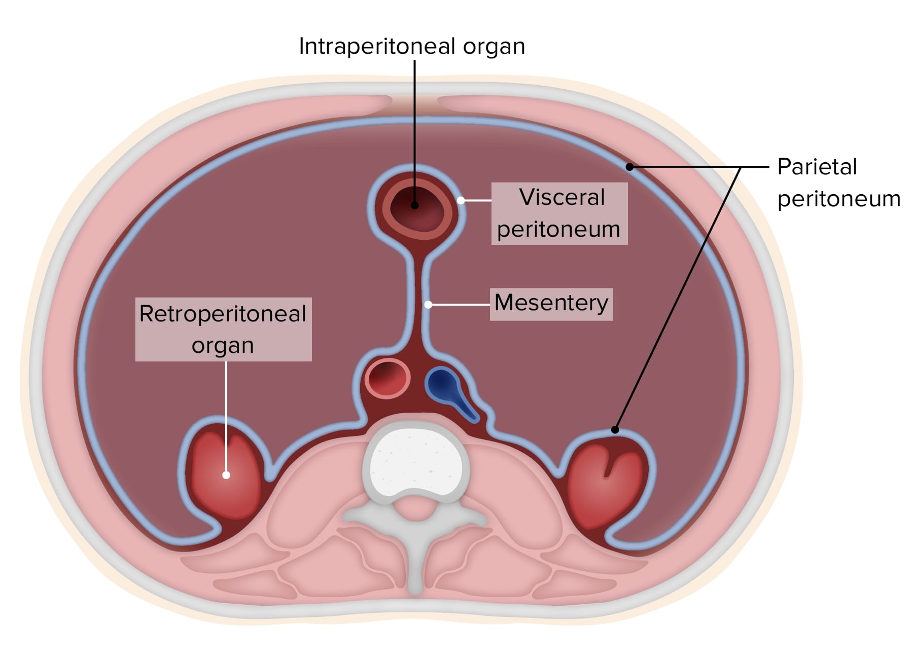
00:01 So in this lecture we will be discussing extraluminal air. 00:04 This is also known as free air, which you may have heard being discussed. 00:08 So where are the different locations of extraluminal air? It can be intraperitoneal. It can be retroperitoneal. 00:16 It can be air within the bowel wall or it can be air within the biliary system. 00:21 So free intraperitoneal air is also called pneumoperitoneum. 00:26 Free retroperitoneal air is also called pneumoperitoneum. 00:31 Retroperitoneal free air is rarely the result of blunt or penetrating trauma. 00:36 Post surgical free air is normal and can be seen for a maximum of about 7 days post surgery. 00:45 So if the patient is not a post surgical patient and they don't have retroperitoneal air due to trauma then this is actually a surgical emergency. 00:53 Any pneumoperitoneum or retroperitoneum is a surgical emergency because it represents perforation of a loop of bowel. 01:00 Free air is best evaluated on CT but it can be seen on radiographs and it should not be missed. 01:06 So it's important to know what free air looks like on a radiograph. 01:09 What are some radiographic signs of pneumoperitoneum? You can see air under the diaphragm and that's really only seen in the upright position. 01:19 You can see both sides of the bowel wall. 01:22 This is called Rigler sign and this can be seen in any position and you can see visualization of the falciform ligament which is also called the football sign and this is seen most often in the supine position. 01:33 Just a reminder that when two structures of the same density are next to each other, they cannot be differentiated on a radiograph. 01:41 Do you remember the name of the sign? This is called the silhouette sign. 01:45 So free air under the diaphragm. 01:48 You can see on this upright chest x-ray that there's a free air under the right hemidiaphragm and you can see this because of the adjacent soft tissue density which is liver down here. So adjacent to the soft tissue density of the liver when you have any bit of air, you can see the air, you can also differentiate it from the adjacent lung because you can see the line right here which represents diaphragm. 02:10 This is really seen only on an upright view because gravity will raise the air superiorly and place it under the diaphragm. 02:17 You can also do a left lateral decubitus film and this is done in patients who cannot stand up or sit upright and it raises the air to the right side adjacent to the liver. 02:27 So again, you see here, this is the patient that's lying on their left side down. 02:31 This is the left side, here are the ribs, so this is the patient?s chest and then here is free air, right adjacent to the soft tissue density of the liver. 02:40 The Rigler sign is visualization of both sides of the bowel wall. 02:46 This is an important sign that seen in free air and the bowel wall is normally not seen because it's adjacent to surroundings soft tissue density of the same density. 02:55 So the bowel wall adjacent to say the liver will up here to be of the same density and you normally wouldn?t see the bowel wall. 03:00 However, if you have air on both sides of it, you now can see the bowel wall. 03:04 So this is an example of the Rigler sign. 03:08 You can see, this is from free intraperitoneal air due to a perforated gastric ulcer and you can see here, this represents the bowel wall and you can see air within the bowel and then you can see air outside of the bowel. 03:21 So this is an important sign representing free air. 03:24 Let's just review with the falciform ligament is. 03:26 It's actually a portion of the peritoneum extending from the anterior surface of the liver to the anterior abdominal wall. 03:33 So right here. It contains the obliterated umbilical vein which is called the ligamentumteres. 03:40 So let's take a look at this case which relates to the falciform ligament. 03:44 What do you see on here? So this right here, is the falciform ligament. 03:57 You can see its extending all the way down from top to bottom and then you can see multiple loops of bowel with bowel wall visible or the Rigler sign. 04:06 This is the CT image of the same patient. 04:11 You can see the falciform ligament attaching to the anterior abdominal wall and then you can see free air on both sides of the falciform ligament. 04:19 So this is an example of the football sign. 04:21 You can also see scattered foci free air, surrounding the liver here as well as over here in the left side of the abdomen. 04:31 So this is an example again of the football sign and another important sign to remember with free air. 04:38 This is seen best on a supine radiograph. 04:47 Let's take a look at this image. Is this the Rigler sign that you're looking at? So this is actually a pitfall. An important pitfall not to make a mistake for. 05:03 You don't wanna mistake multiple dilated loops of the bowel that are adjacent to each other and consider that free air. 05:10 So this is a patient that has multiple dilated loops of bowel probably a small bowel obstruction and you can see bowel wall adjacent to this air within the bowel. 05:20 So you would think that this could represent the Rigler sign. 05:23 If there's any kind of confusion the next step would be to perform a CT to see if there really is free air or rather this represnt a small bowel obstruction. 05:33 So what are some radiographic signs of pneumoperitoneum? Again, this is free air within the retroperitoneal space. 05:38 So air will actually outline the retroperitoneal structure such as the psoas muscles or the kidneys and it may traverse the diaphragmatic hiatus and go up into the chest causing pneumomediastinum. 05:50 This is an example of retroperitoneal air outlining the IVC. 05:53 And it also outlines a portion of aorta. 05:56 So this right here is the location of the IVC. 05:57 You can see free air surrounding that and you see free air here retroperitonealy surrounding the aorta as well and up in the left upper abdomen.
About the Lecture
The lecture Extraluminal Air: Pneumoperitoneum & Pneumoretroperitoneum by Hetal Verma, MD is from the course Abdominal Radiology.
Included Quiz Questions
Which of the following is NOT true regarding pneumoperitoneum?
- It is always a surgical emergency.
- It is associated with the Rigler sign.
- It is associated with the football sign.
- It is best seen on an upright abdominal or chest film under the diaphragm.
- It can be seen up to 1 week postoperatively.
A 22-year-old man underwent an appendectomy 2 days ago. He now complains of vague pain in his abdomen with a distended abdomen. Free air can be best evaluated with which of the following diagnostic methods?
- CT scan of the abdomen
- MRI of the abdomen
- Ultrasound of the abdomen
- X-ray
- Endoscopy
A patient presenting with pain in abdomen is sent for a CT abdomen as a part of the diagnostic evaluation. The CT shows air surrounding the inferior vena cava and a portion of the aorta. What does this finding signify?
- Pneumoretroperitoneum
- Pneumoperitoneum
- Inferior vena cava rupture
- An artifact
- Inferior vena cava thrombosis
Author of lecture Extraluminal Air: Pneumoperitoneum & Pneumoretroperitoneum

Hetal Verma, MD
Customer reviews
5,0 of 5 stars
| 5 Stars |
|
5 |
| 4 Stars |
|
0 |
| 3 Stars |
|
0 |
| 2 Stars |
|
0 |
| 1 Star |
|
0 |
