Playlist
Show Playlist
Hide Playlist
Complete Picture of the Cardiac Cycle – Cardiac Cycle


-
Slides CardiacCycle CardiacPhysiology.pdf
-
Download Lecture Overview
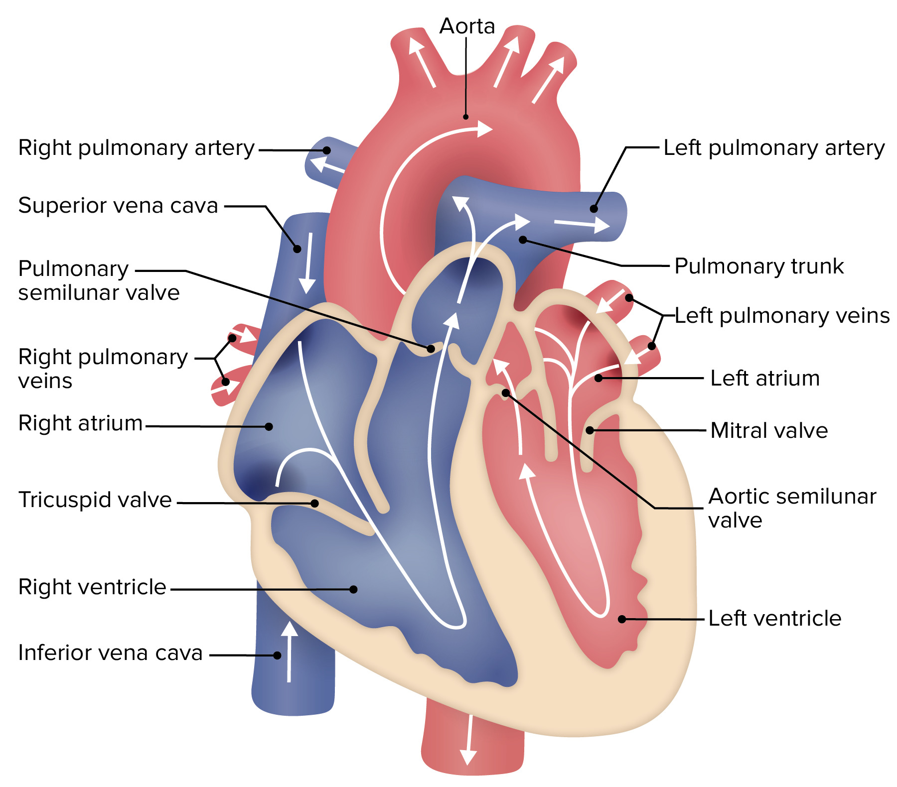
00:01 Now, we're going to connect two big points here. 00:07 We've talked about the cardiac cycle and we talk about the cardiac cycle over time. 00:14 Why? Because there is a time component to the ECG. 00:18 We went through all of these different items, such as there’s an R-R interval, there's a P-R interval, there's a Q-T interval. 00:25 They'll have specific, specific times. 00:29 That helps us understand how the heart works in sequence. 00:35 But to really understand the function of the heart, we’re going to have to plot the graph in a little different manner. 00:43 To do this, we are going to flip two of the axes. 00:48 We’re going to remove time out of the equation and we’re going to put this left ventricular volume from being on its own in the cardiac cycle, we’re going to slide it over to the x-axis. 01:00 And on the y-axis, all we’re going to have is left ventricular pressure, which is seen here in the solid red line above. 01:13 So, let's now talk through what things happen in a pressure-volume curve. 01:20 Make sure you follow along this very carefully, so that you can link everything back to the cardiac cycle because I've taught this for many years and I know that it's very easy to understand the cardiac cycle with all of its different components. 01:36 You know you have a contraction. 01:37 You have relaxation. 01:39 You have filling. 01:39 You have all these things that come in sequence. 01:43 Some people don't see the sequence on a pressure-volume curve as quickly, but as soon as you do, you will now have insight and ways that you can now express changes in the cardiovascular system that you don't have the ability to express with the cardiac cycle. 02:01 So, if we plot left ventricular volume on the X, left ventricular pressure on the Y, we're going to start at the end systolic volume and start with filling. 02:12 And that's denoted as A on your figure. 02:16 So, the filling phase is along the bottom, then you have isovolumic contraction, then you have this rapid and reduced ejection and then isovolumic relaxation. 02:30 So, you have all of the same components there, but now they're just located in a loop rather than over time. 02:40 So, what happens at the various spots. 02:44 Well, the mitral valve opens at A, so now you can start to fill the left ventricle. 02:51 You can have rapid filling, reduced filling, and the atrial kick. 02:59 Then the mitral valve closes and that’s denoted as B. 03:04 And interestingly, this A and B are important points. 03:09 This corresponds to your left ventricular end-systolic volume at A and left ventricular end-diastolic volume at B. 03:18 You subtract those two and you get stroke volume. 03:23 Then you have an isovolumetric change, and that is seen by that vertical line up. 03:31 So, now, you're going from B to C. 03:34 And as soon as you hit C, you've now eclipsed the pressure of the aorta. 03:41 So, now, you can open up the aortic valve. 03:43 Once aortic valve is open, you have a rapid ejection followed by a reduced ejection, then the aortic valve closes, and that’s denoted here by D. 03:56 Between D and A is simply isovolumic relaxation. 04:03 So, you see how you can express the entire cardiac cycle on this one little curve.
About the Lecture
The lecture Complete Picture of the Cardiac Cycle – Cardiac Cycle by Thad Wilson, PhD is from the course Cardiac Physiology.
Included Quiz Questions
Which of the following is used to express stroke volume?
- End diastolic volume (EDV) - End systolic volume (ESV)
- End systolic volume (ESV) + End diastolic volume (EDV)
- Cardiac output × Heart rate
- End systolic volume (ESV) - Cardiac output
- End systolic volume (ESV) ÷ End-diastolic volume (EDV)
Which stage of the cardiac cycle do the atrioventricular valves open?
- Ventricular filling
- Atrial filling
- Isovolumetric contraction
- Ventricular ejection
- Atrial contraction
Author of lecture Complete Picture of the Cardiac Cycle – Cardiac Cycle

Thad Wilson, PhD
Customer reviews
5,0 of 5 stars
| 5 Stars |
|
5 |
| 4 Stars |
|
0 |
| 3 Stars |
|
0 |
| 2 Stars |
|
0 |
| 1 Star |
|
0 |
