Playlist
Show Playlist
Hide Playlist
Anatomy of the Dura

-
Slides Introduction to CNS Infection.pdf
-
Download Lecture Overview
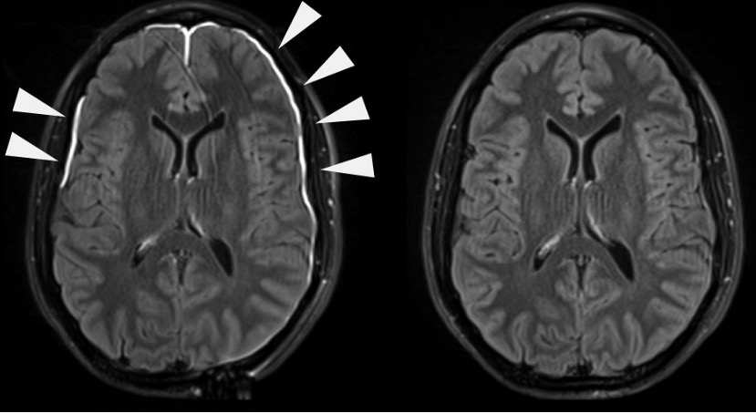
00:00 Let's start with some anatomy. In order for an infection to occur, an organism has to get through the body's defenses and one of the most robust defense mechanisms is sheltering of the brain and the nervous system from the outside world. What does that? Well, when we move from the outside world into the brain, we got to pass through the scalp. And the scalp stands for first the skin. Slightly deeper we get to the connective tissue, that's the C. The A is called the aponeurosis, a thick fascial layer. It is just beneath the skin and connective tissue and is a thick fascia. Aponeurosis are called the galea aponeurotica. Beneath the galea is loose connective tissue, a very thin layer of loose, L, connective tissue. And then the periosteum. And together those 5 layers make up what we call the scalp. Beneath the scalp is the bone and we know that calvarial or cranial bone is composed of an outer cortical bone and intercortical bone and in between that is a diploic space, soft tissue or soft area of bone. 01:08 And then beneath the bone is the outer covering of the brain, which we call the meninges. 01:12 First, we see the dura mater and there are reflections in the dura where the venous sinuses occur, and you can see that in the middle part of this picture. Beneath the dura, we have the arachnoid and the subarachnoid space, a potential space where blood vessels lie, and then the pia mater. And together, the pia, arachnoid, and dura make up what we call the PAD or the meninges. And deeper still, we get to the brain or the intraparenchymal compartment of the brain tissue. So let's take a closer look at this and each compartment of the nervous system and brain. Again, we have the dura, the arachnoid, the pia mater, and the brain and we're going to look at infections that happen that are concentrated in each of those areas. 02:00 Meningitis is defined as inflammation of the meninges with preserved brain function. 02:04 The three meningeal layers are all affected. 02:07 When the brain parenchyma is affected, we can think of encephalitis which is inflammation of the brain tissue due to viral pathogens; or cerebritis, the inflammation of brain parenchyma due to bacterial pathogens or abscess formations. 02:19 So, again, we're going to learn about three types of infections, meningitis, encephalitis, and cerebritis. 02:26 When we think about the dura and look at the dura, the infection that we're going to look into and focus on will be meningitis. 02:34 Meningitis is an infection that primarily affects the dura matter. 02:38 That's where we see it on imaging, that's where we evaluate it when we're looking at patients and this is where many of the symptoms come from. 02:45 In the level of the arachnoid in pia, the infection that occurs primarily in that area is encephalitis. 02:52 And when we think of encephalitis we're going to think of a problem that's in the arachnoid, the pia mater, and that subarachnoid space, that potential space in between those two layers. 03:02 The last layer is the brain, the intra-parenchymal tissue. 03:05 And an infection that occurs in that area is cerebritis. 03:09 Cerebritis is an infection in the brain parenchymal compartment, and that will account for both cerebritis and brain abscesses. 03:18 So again, we're going to learn about three types of infections, meningitis, encephalitis, and cerebritis; and when we think about those, we're going to think about those infections affecting the dura mater, the arachnoid and subarachnoid space in pia, and the brain tissue.
About the Lecture
The lecture Anatomy of the Dura by Roy Strowd, MD is from the course CNS Infections.
Included Quiz Questions
The space between the arachnoid mater and pia mater is called the...?
- ... subarachnoid space.
- ... epidural space.
- ... extradural space.
- ... subdural space.
- ... periosteal space.
Author of lecture Anatomy of the Dura

Roy Strowd, MD
Customer reviews
5,0 of 5 stars
| 5 Stars |
|
5 |
| 4 Stars |
|
0 |
| 3 Stars |
|
0 |
| 2 Stars |
|
0 |
| 1 Star |
|
0 |
