Playlist
Show Playlist
Hide Playlist
Fertilization to Bilaminar Embryo


-
Slides 01-02 Fertilization to Bilaminar Embryo.pdf
-
Download Lecture Overview
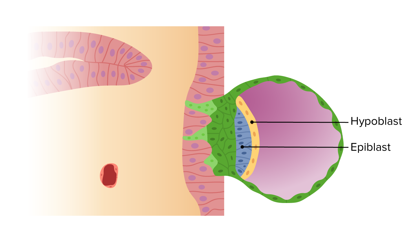
00:01 In this talk, we will follow the progress of the earliest steps in embryology, from fertilization to formation of the bilaminar embryo. 00:09 We're gonna pick up right here at the very moment after fertilization has occurred. 00:15 We can see that the spermatozoon has just entered the oocyte. 00:20 It has punctured through the protective barrier of cells and the zona pellucida to reach the oocyte and its cytoplasm. 00:28 As soon as a single spermatozoon has entered the cytoplasm of the egg, it sets off a chain reaction of events that prevent additional spermatozoa from making it in. 00:38 After the sperm penetrates the oocyte, the oocyte completes meiosis II. 00:43 This step is important because it is responsible for forming the ovum and the second polar body. 00:50 At that point, the one male pronucleus consolidates inside the cytoplasm and it begins to associate with the female pronucleus. 01:00 Once the two pronuclei approach each other, a mitotic spindle forms between them. 01:06 Now, these two fuse and come together and we have got the genetic material, making up a brand-new and distinct individual. 01:16 At this point, we have the diploid complement of chromosomes that can be used as the blueprint for all the subsequent formations and changes that will bring about the human being that can grow from this single cell. 01:30 As these nuclei have formed, a series of events can begin that have been on hold for quite a while in the egg. 01:37 In particular, mitotic division can start taking place again and the chromosomes can go through the whole process of metaphase, anaphase, telophase, and produce a subsequent generations of cells that are genetically similar, in fact, identical to the progenitor cells. 01:54 Roughly 30 hours after fertilization, the first split is going to occur and have two daughter cells to take the place of the single zygote. 02:03 Now, at this point, subsequent divisions are going to occur and the resulting cells will not grow appreciably. 02:10 In fact, they'll take up as much space as the original, single-celled zygote did. 02:15 So, they will get smaller and smaller even as they become more numerous. 02:19 At this point, we're gonna refer to the entire assemblage as a morula, a collection of cells, and we're gonna take a look at the nine-cell stage, roughly two and a half days into development. 02:30 At this time, we've got something about the same size as a zygote and it's gonna consist of a group of cells clustered in the center, the inner cell mass, and the surrounding outer cell mass. 02:43 During this time, the protective barrier that existed around the developing zygote and morula, the zona pellucida begins to break down and fluid begins to permeate into the spaces between these cells. 02:56 Thereafter, we're gonna go a bit further to a stage of development called the blastocyst. 03:02 The fluid that is moved into the spaces between the cells of the morula creates a cavity called a blastocyst cavity and the entire assembly of fluid and cells is referred to as the blastocyst. 03:15 Now, the early blastocyst has roughly 58 cells and it will continue to develop to roughly the 107-cell stage after five days which is gonna be called the late blastocyst. 03:26 It looks about the same with the fluid-filled cavity surrounded by cells on the outside and a cluster of inner cells pushed off to one pole of the blastocyst. 03:37 The inner cell mass and outer cell mass will form very distinct structures. 03:43 As we proceed from the morula to the blastocyst and further on into development, we'll see that the outer cell mass transitions into a set of structures called the trophoblast and the inner cell mass is going to become the embryoblast and the embryoblast is what will generate all the tissues of the actual embryo and fetus. 04:03 Whereas the trophoblast will become the supporting tissues of the placenta and the umbilical cord. 04:09 Now, let's take a look at how development proceeds from the blastocyst into the bilaminar embryo and during this time, we have the embryo moving into the uterine wall from day 7 to 15. 04:25 Initially, the inner cell mass, which will form the embryoblast, subdivides into a thin layer of cells in contact with the blastocyst cavity called the hypoblast and a taller group of cells further away from the blastocyst cavity called the epiblast. 04:41 The epiblast and hypoblast cells have distinctive fates. 04:46 The hypoblast cells are going to be proliferating to surround the blastocyst cavity. 04:52 They are small and cuboidal. Whereas the epiblast cells grow taller and taller and they're going to be the cells that actually create the entirety of the embryo from this stage on. 05:03 Now moving into the uterus, the developing blastocyst is going to have these epiblast cells and hypoblast cells to a few different things. 05:14 In particular, a small cavity is going to form in the epiblast cells called the amniotic cavity. 05:20 Now, this tiny little pinpoint of a cavity may not look like much right now, but it will be the cavity that surrounds the entirety of the embryo and fetus, forming the water that supports and nurtures it throughout developments. 05:35 The amniotic cavity will eventually surround the entire fetus. 05:39 As we go further and further into the uterus, the hypoblast cells are going to proliferate and spread out across the blastocyst cavity. 05:48 Once they've completely done so, an extended, what sometimes called Heuser's membrane throughout that space, that entire cavity will transition to become primary yolk sac. 05:59 Moving a little further along, the primary yolk sac and embryo become separated from the surrounding trophoblast cells by a distinctive set of cells called the extraembryonic mesoderm. 06:13 This extraembryonic mesoderm will not contribute anything of substance to the actual embryo but allows it to develop fully inside the uterus and separates it from the placenta. 06:25 This happens because some cavities are going to start appearing inside the extraembryonic mesoderm and these cysts are little cavities are going to form, fuse, and become what's known as the extraembryonic coelom, a space that forms outside of the developing embryo and as it enlarges and completely wraps around the yolk sac and the embryo, it will form what's called the chorionic cavity. 06:51 Initially, the chorionic cavity will surround the embryo and only much later be replaced by the expanding amniotic cavity. 07:00 So, as we move a little further along, the expansion of the chorionic cavity pinches the yolk sac in such a way that a portion of it moves away from the embryo towards the opposite pole of the developing embryoblast. 07:13 The part of the yolk sac that remains in contact with the embryo is going to be distinct and it's going to form what's called the definitive yolk sac. 07:23 The portion that moves away is going to form remnants of the primary yolk sac called exocoelomic cysts. 07:30 Now the secondary or definitive yolk sac stays in contact with the epiblast cells that are developing into the embryo and have an ever-enlarging amniotic cavity within. 07:42 Now here, as the chorionic cavity and the placenta has grown larger and larger, we can see that the extraembryonic mesoderm is lining the developing placenta and chorionic cavity and is only connected to the developing embryo by a single small connecting stalk. 08:02 That connecting stalk is what connects the developing bilaminar embryo to the developing placenta and that is what will become the umbilical cord. 08:12 Thank you very much for your attention and I'll see you during our next talk.
About the Lecture
The lecture Fertilization to Bilaminar Embryo by Peter Ward, PhD is from the course Early Development and the Organogenic Period. It contains the following chapters:
- Fertilization to the Bilaminar Embryo
- Development from the Blastocyst into the Bilaminar Embryo
Included Quiz Questions
During fertilization, the spermatozoon enters the ovum by puncturing what non-cellular structure?
- Zona pellucida
- Zona fasciculata
- Zona gale
- Zona radiata
- Zona reticularis
What is a morula?
- 16-cell mass
- 2-cell mass
- 8-cell mass
- 4-cell mass
- 58-cell mass
Which of the following refers to the cavity without cells, that is formed by the permeation of fluid into morulae?
- Blastocele (blastocyst cavity)
- Blastocyst
- Morulae
- Zygote
- Trophoblast
Which structure gives rise to the placenta?
- Trophoblast
- Embryoblast
- Zona pellucidum
- Inner cell mass
- Corona radiata
What is Heuser's membrane?
- Hypoblast cells lining the blastocyst cavity
- Epiblast cells lining the amniotic cavity
- Proliferating epiblasts within the blastocyst
- The inner lining of the morula
- The outer layer of cells of the blastocyst
Which of the following will give rise to the umbilical cord in the embryoblast?
- Connecting stalk
- Primitive yolk sac
- Trophoblast
- Extraembryonic coelom
Author of lecture Fertilization to Bilaminar Embryo

Peter Ward, PhD
Customer reviews
5,0 of 5 stars
| 5 Stars |
|
6 |
| 4 Stars |
|
0 |
| 3 Stars |
|
0 |
| 2 Stars |
|
0 |
| 1 Star |
|
0 |
I loved this lecture! Now I truly understand the first two weeks of embryo development! Thanks Dr.Peter !
MA SHA ALLAH.. It was great and easy to understand I am glad
perfect and it was easy to follow . i like the way of questions
Highly organized and well structured lecture. He clearly sets out events on a timeline and explains what happens. Engaging.
