Playlist
Show Playlist
Hide Playlist
Standard 12 Lead ECG
-
Slides Learning to Read an ECG.pdf
-
Download Lecture Overview
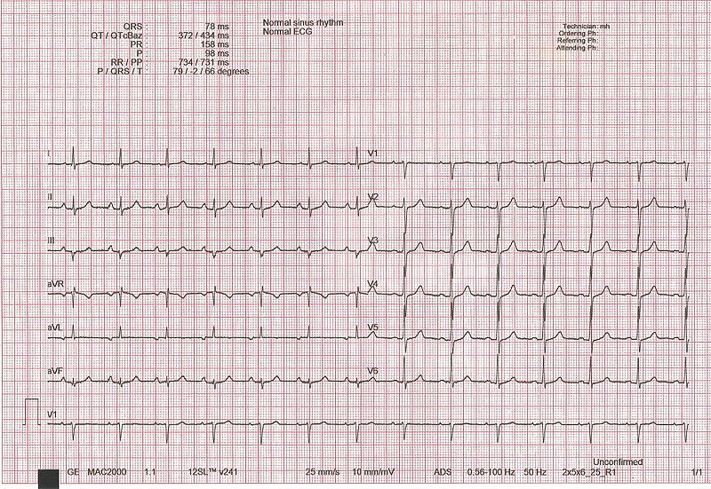
00:01 So, it turns out that the - by convention over the years, the EKG has come to have 12 different leads. 00:09 That is 12 different points that are looking at the wave of depolarization going down through the heart. 00:16 And the earliest ones from Einthoven were so-called leads I, II and III. 00:22 So, these leads are all designated by the points on a compass, 0 to 360 degrees. 00:29 Lead I is over here and it's looking in at the heart from the left point - like the left arm in. 00:40 And that is designated as zero degree. Lead II is looking from here; that's +60 degrees. 00:47 And lead III is looking from here; that's +120 degrees. 00:53 Well over the years, there have been additional leads added, and that these are the so-called augmented leads. 01:00 Leads aVR, aVL and aVF. Lead aVF is looking from straight down below; that's +90 degrees. 01:07 aVL is looking from -30; that's up here. 01:11 And aVR is looking from +210 from right over here on the right arm. 01:16 These are all looking in a frontal plane just as if we were a flat plane of - that the body was standing in. 01:25 So here we see again the leads I said before, the Einthoven leads. 01:29 Lead I coming in from the left arm; that's zero degree. Lead II, +60 degrees coming from here. 01:35 And lead III, 120 degrees plus. 01:39 We also have leads that are cutting the heart right down in a section just as if it were right through the body like this. 01:46 Those leads are placed on the chest out here. 01:49 Here's lead I, lead II, III and IV starting at the right sternal border and working around to the axilla. 01:56 And they give you views of the depolarization wave in a plane and that's V1, 2, 3, 4, 5 and 6. 02:07 And all of these leads, well, the ones we said before, lead I, II and III, aVR, aVL and aVF, V1, 2, 3, 4, 5 and 6 are all printed out on a piece of paper or appear on a computer screen for interpretation.
About the Lecture
The lecture Standard 12 Lead ECG by Joseph Alpert, MD is from the course Electrocardiogram (ECG) Interpretation.
Included Quiz Questions
Which of the following statements regarding the precordial ECG lead system is FALSE?
- All of the leads are placed on the same side of the sternum.
- There are 6 precordial leads.
- V1 and V2 are the most anterior.
- V5 and V6 are the most lateral.
- Precordial leads give information in the transverse plane.
Customer reviews
3,0 of 5 stars
| 5 Stars |
|
1 |
| 4 Stars |
|
0 |
| 3 Stars |
|
1 |
| 2 Stars |
|
0 |
| 1 Star |
|
1 |
This is a great lecture. Shame about the previous reviews. If you didnt study maths in school, you may not have understanding of vectors. This is a shame.
Should have gone into way more detail about the leads
I liked the summary, it lacked some key information like what positive and what is negative and why, what is an unipolar and bipolar lead, etc. But it's fine, you'll probably need some basic electrophysiology knowlegde to not get distracted by why things are how they are.


