Playlist
Show Playlist
Hide Playlist
Epidural Hemorrhage – Intracranial Hemorrhage (ICH)

-
Slides 21 IntracranialHemorrhage HeadNeckAnatomy.pdf
-
Download Lecture Overview
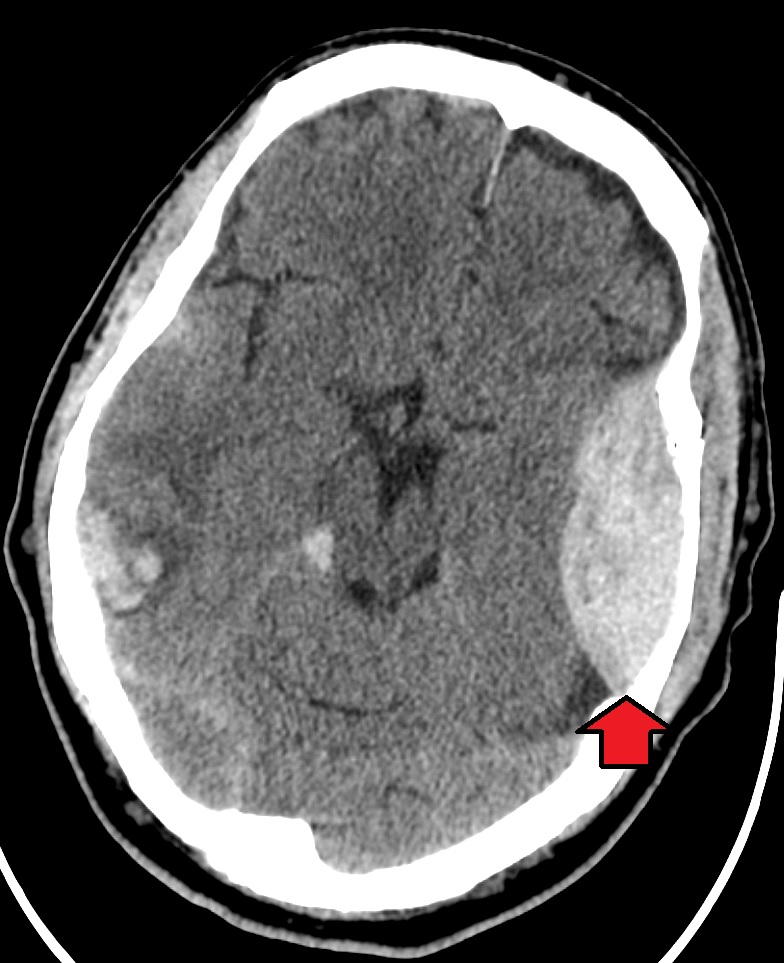
00:01 Let’s begin now and focus on the epidural hemorrhage. We need to understand what the vascular source of this type of hemorrhage comes from. The vascular source of an epidural hemorrhage or a bleed would be from the branches of the middle meningeal artery. 00:23 The middle meningeal artery is a branch of the maxillary artery that enters the skull through the foramen spinosum. As we take a look here, we can see the middle meningeal artery and we see its branching pattern. This is an anterior branch. This area here, the middle meningeal artery is coursing deep to the skull within the cranial cavity. It will groove the skull at this particular point internally. This area of the skull is referred to as the pterion. This is one of the thinnest portions of the skull on this side. So a blow here can fracture the skull. 01:01 Then this is the posterior division that has a more posterior course as we see here. 01:07 So, the most common cause of an epidural hemorrhage is indeed a traumatic cause. 01:12 So there is a severe blow to the side of the skull usually in the area of the pterion that’s sufficient to fracture this region of the skull. Then the fractured bone gets pushed in internally and then can sever one of the branches of the middle meningeal artery. In the case of the pterion in this area here, that would be the anterior division of your middle meningeal artery. 01:42 Here, we see a section through the skull demonstrating an epidural hematoma. 01:50 Here, we can see the fracture of the skull right in through here on the lateral side. 01:58 As a result of that fracture, the middle meningeal artery has been severed. Because this is under high pressure, you’ll get a separation of the dura away from the skull. You see the fairly large substantial bleed here within the cranial cavity. The layer of the dura that is separating from the skull will be the periosteal layer. As a result of this separation of the periosteal layer of the dura mater from the skull, the hematoma creates a pathologic space which is a bleed into the epidural space. This is normally not present except in this form of pathology. A characteristic of an epidural hematoma is its shape. As we can appreciate in this view, the epidural hematoma will produce a lens-shaped appearance. 03:00 A very important consideration of an epidural hematoma because of the rapid bleed because this artery is under high arterial pressure, the hematoma can and usually does cause a shift of brain structures that will then deviate to the opposite side. So you can see a shift here of this side of the image being shifted over to the right side of the image because of this high pressure of this space-occupying lesion. In addition, the increase in intracranial pressure can cause a herniation of a couple of brain structures. Here, we see herniation of the uncus in through here, through an opening in the tentorium cerebelli. 03:58 Then down inferiorly here where we have the medulla and the spinal cord transition occurring, you can see herniation of the cerebellum through the foramen magnum. These are very, very serious herniations and can result in the death of a patient.
About the Lecture
The lecture Epidural Hemorrhage – Intracranial Hemorrhage (ICH) by Craig Canby, PhD is from the course Head and Neck Anatomy with Dr. Canby.
Included Quiz Questions
The middle meningeal artery is a branch of which of the following?
- Maxillary artery
- Vertebral artery
- Subclavian artery
- External carotid artery
- Internal carotid artery
Which of the following locations is most susceptible to epidural hematoma formation?
- Pterion
- Nuchal line
- Semispinalis capitis
- Condyloid fossa
- Temporal line
Which areas does an epidural hematoma create a pathological space between?
- Skull and periosteal layer of dura mater
- Dura mater and arachnoid mater
- Skull and pia mater
- Pia mater and periosteal layer of dura mater
- Dura mater and pia mater
Author of lecture Epidural Hemorrhage – Intracranial Hemorrhage (ICH)

Craig Canby, PhD
Customer reviews
5,0 of 5 stars
| 5 Stars |
|
5 |
| 4 Stars |
|
0 |
| 3 Stars |
|
0 |
| 2 Stars |
|
0 |
| 1 Star |
|
0 |
