Very helpful
By Alaena A. on 19. October 2021 for Female Pelvic Anatomy
Very helpful overview of normal anatomy as seen on US, CT, and MRI
Requires viewer to do their own learning on Google.
By Alaena A. on 08. October 2021 for Benign Liver Disease: Hepatic Steatosis, Cavernous Hemangioma and Hepatic Cyst
organization of the MRI section is confusing. It would have been better to show the definitions and the pictures at the same time...instead of using terms not previously introduced...and then switching to the definitions, but now the viewer can't see the image which demonstrates the terms. Instead, I had to look up the terms on my own and then I could better understand the lecture. I also had to spend time going back and forth with the slides and transcript/video to better understand. In short, the organization of this section could have been done better.
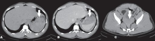
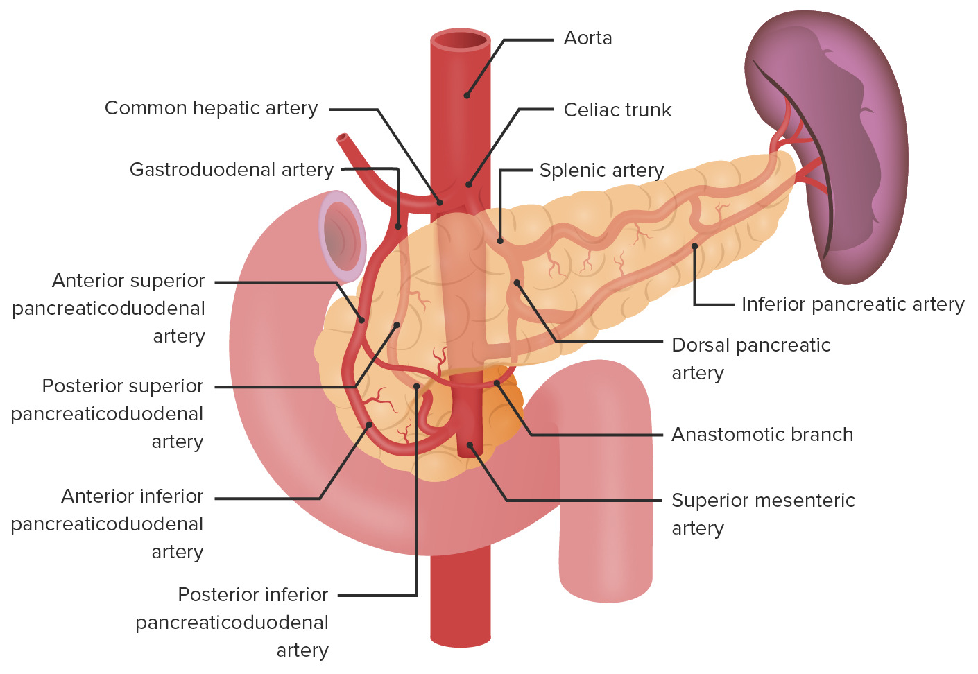
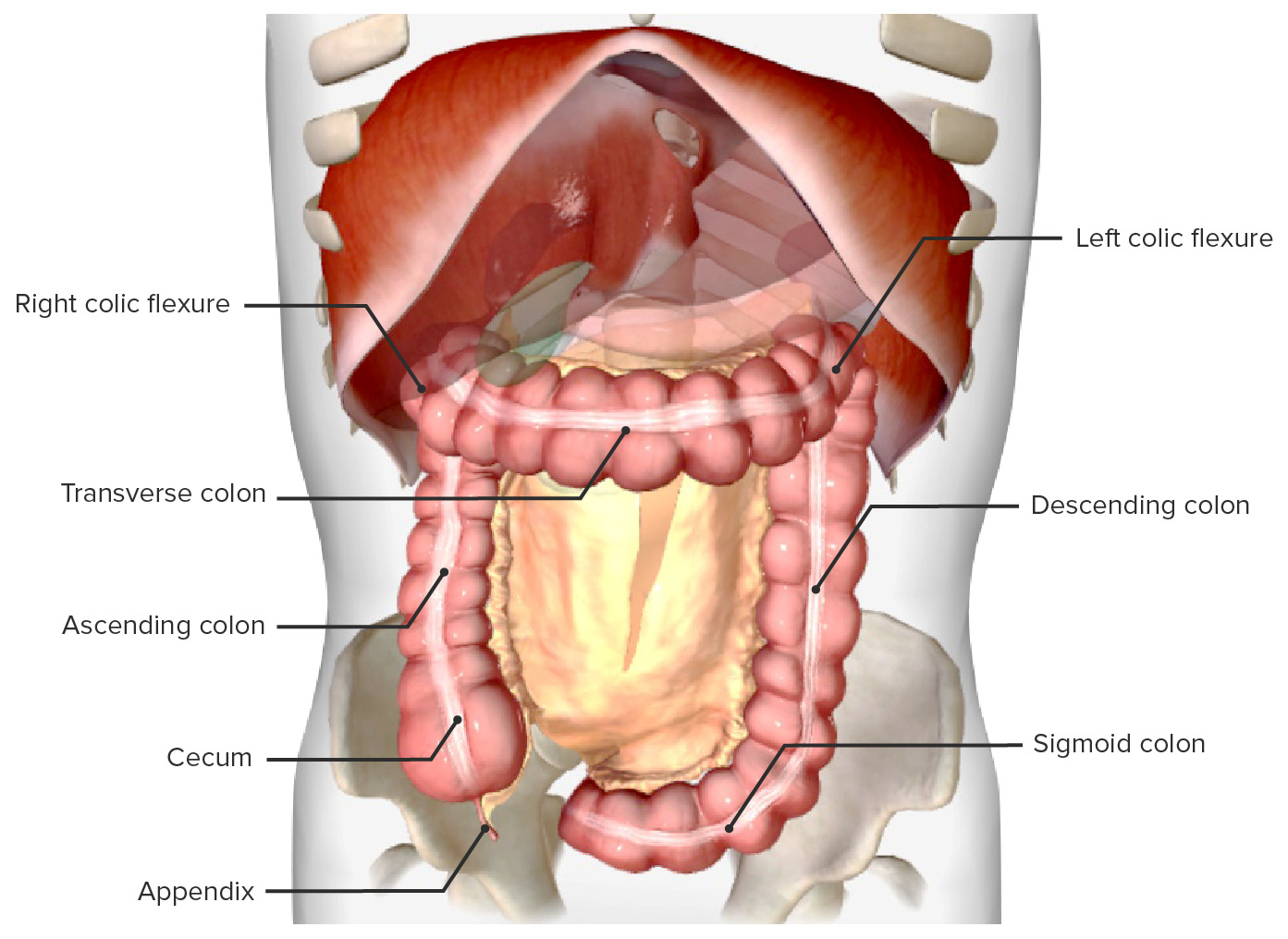
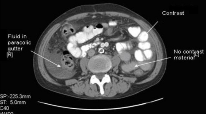
tYpes of calcifications
By Mbanzabugabo J. on 03. October 2021 for Abdominal Radiology
i was impressed the way abdominal is well summarize
Thank you very much, it is amazing
Lecture is great
By Irfan A. on 26. March 2021 for Abdominal Radiology
Like it very much, like it very much, like it very much
excellent, I would recommend this course, therefore rating 5 stars
By rosemarie m. on 03. September 2020 for Abdominal Radiology
Excellent, I trained in India, and her voice is good. Am I with Leipzig- Brahms city - been there, also when it was GDR. Been down the road to Dresden, remember Trabis on the transit road in 1971.
good
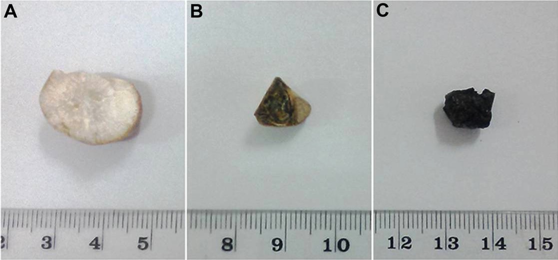
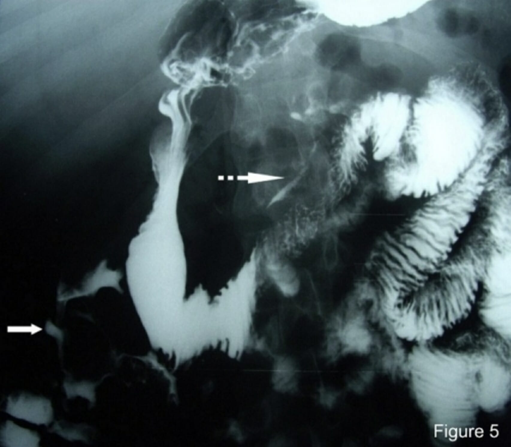
By lk l. on 30. May 2020 for Pancreatic Abnormalities
good and very very goood and thanks i wish i meet you some day
easy and simple and short and most important and clear
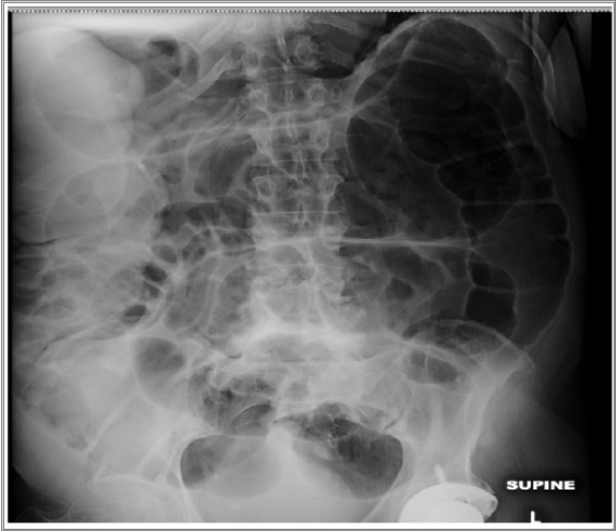
By jk g. on 22. May 2020 for Fluoroscopy: Small Bowel Follow-Through, Tube Study and Barium Enema
good easy and simple and short and most important and clear
Very nice
By Anne V. on 02. May 2020 for Abdominal Radiology
Very well explained! Love the pause suggestions. Prof Verma has a nice and clear voice
Very good!
By Filipa D. on 17. March 2020 for Abdominal Radiology
Very good! Just one thing that i dislike, teacher speaks very fast, because she is reading almost of time, she just speaks slower when she is explaining something for example in a x-ray. So for that reason i could not understand everything at first time when i was watching.
Excellent lecture
By Mathew C. on 22. January 2019 for Abdominal Radiography
Concise and well presented. Delivered all the necessary information without wasting any time
Very clear and contain very interesting informations
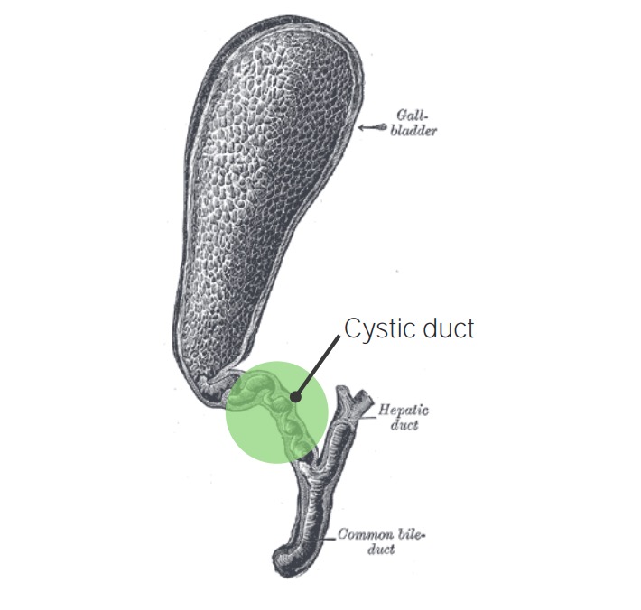
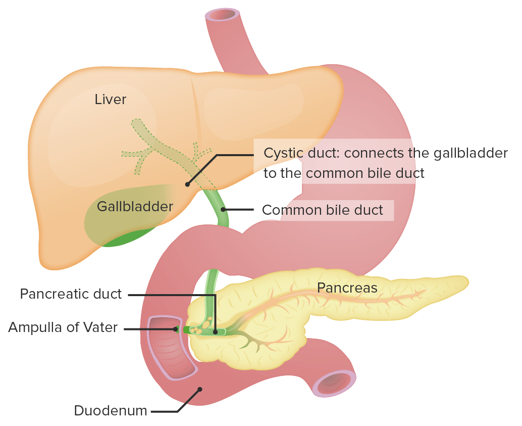
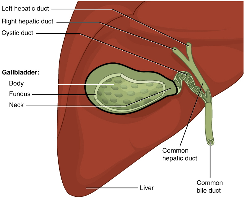
By Louis S. on 17. September 2018 for Gallbladder Abnormalities
All of your lectures about radiology are very clear and contain very interesting informations !!! You're one of the best lectors of Lecturio in my humble opinion. Thanks !!
Excellent course
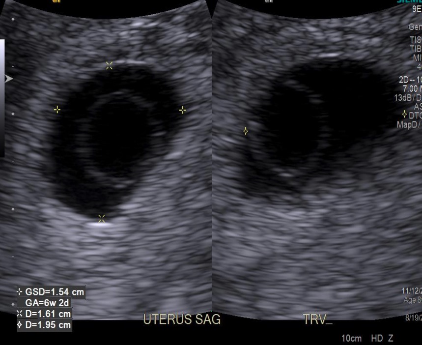
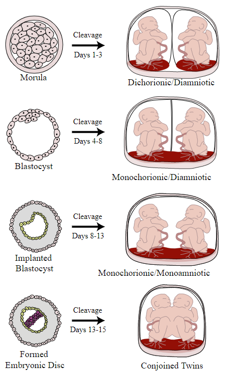
By Okechukwu V. on 25. May 2018 for Heterotopic and Molar Pregnancy
Radiology is no longer just shadows for me. The concepts are well explained in proper clinical contexts.
Congratulations !!
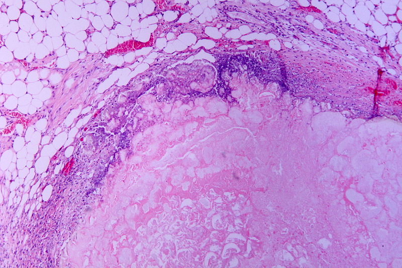
By Louis S. on 23. May 2018 for Hepatic Cirrhosis
Very good lecture !! Doctor Verma teaches with pedagogy and repeats the informations to be we able to better memorize ! Thanks
Great lectures.
By Carmen S. on 16. January 2018 for Abdominal Radiology
Fan tastic lectures.
Now I can understand several topics about imaging.
radiology made simple
By Elona P. on 29. December 2017 for Bladder Carcinoma
i never thought radiology would be so simple! i loved every lesson!
Best lead into the subject..
By Prabhu G. on 01. December 2017 for Abdominal Radiology
Being one scared to go through the subject of radiology.. Your lectures made me familiar with basics made them easy and handy making me love to finish my preparation complete with radiology .. Thank you so much!!
AMAZING ABDOMINAL RADILOGY
By festus s. on 10. July 2017 for Abdominal Radiology
Well presented and very rich abdominal Anatomy. such lectures are very educative. cost effective as there is really no need to jump on the plane.
keep this spirit going.
FESTUS FROM ZAMBIA.