Playlist
Show Playlist
Hide Playlist
Early Gastrointestinal Development

-
Slides 07-41 Endoderm derivatives .pdf
-
Download Lecture Overview
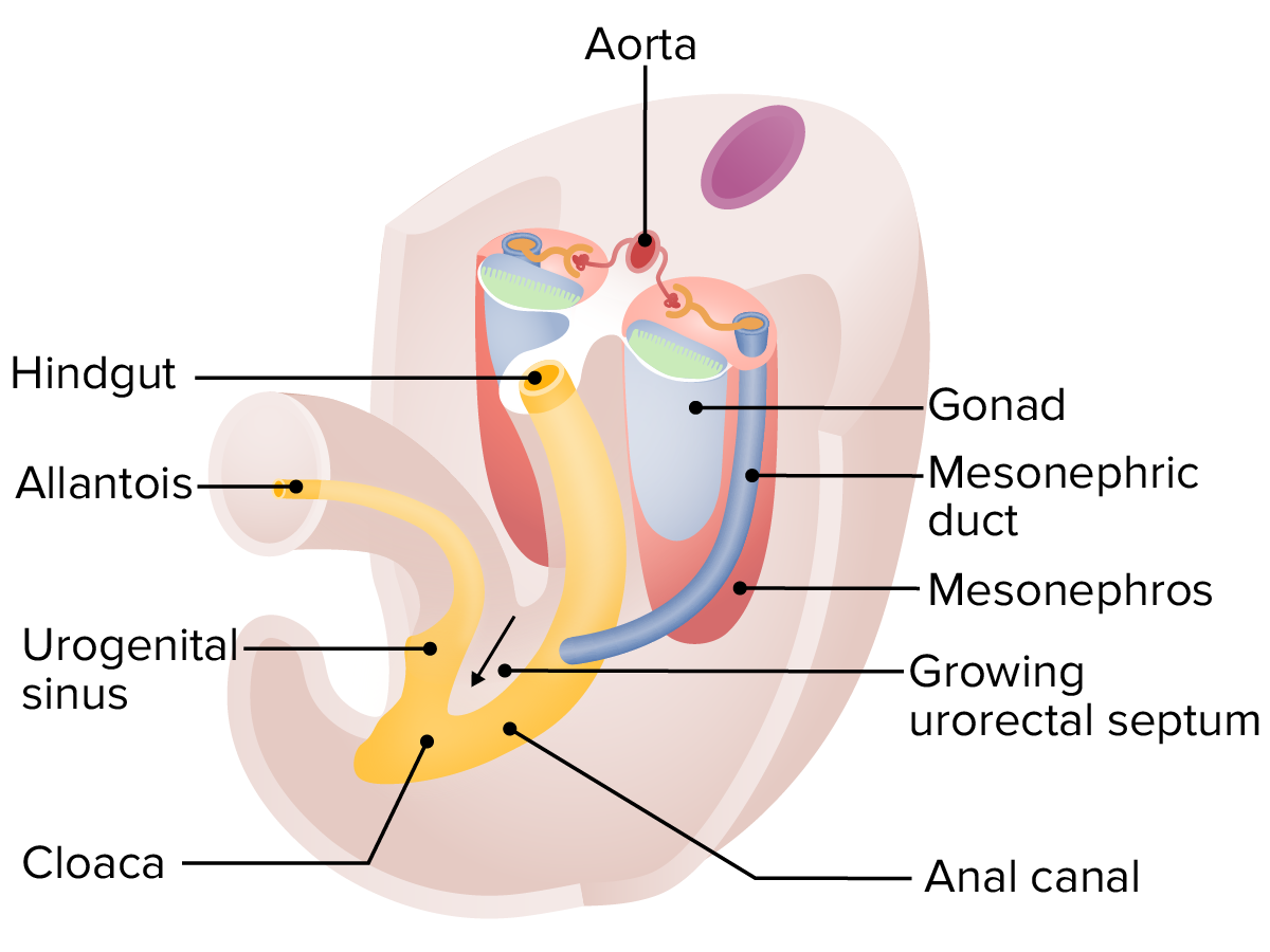
00:01 We will begin discussing how the gastrointestinal system develops by looking at the endoderm and how it folds to create the original gut tube and its associated structures. 00:11 Now, the gastrointestinal system begins as a simple tube from mouth to anus but some very interesting things happen along the way. 00:19 It has to extend, elongate, and develop a variety of glands that are associated with it such as the liver and the pancreas. 00:28 As this happens, the gut actually runs out of space in the abdominal cavity and has to herniate temporarily into the umbilical cord. 00:36 The foregut organs not only grow larger but migrate, shift around, and pull their mesenteries along with them creating some bizarre structures such as the omentum and various mesenteries that connect different parts of the gut tube to the body wall. 00:52 And last but not least, the common chamber for the reproductive, urinary, and digestive tracks, the cloaca, has to be subdivided to keep those systems separate from each other at the time of birth. 01:05 So let’s begin by returning to an earlier stage of development, a roundabout the trilaminar embryo stage as Neurulation is occurring and as the neural folds are moving in to create the central nervous system, the splanchnopleure and its underlying endoderm are folding together to create a gut tube and in the process, extend the yolk sac off of and out of the body wall. 01:30 Now, the body wall is going to move forward the somatopleure surrounding the splanchnopleure and it’s going to form a definitive body wall both in the thorax, abdominal, and later, the pelvic region. 01:43 But as it does so, it needs to allow a little bit of space for two structures to exit the body. 01:48 The first is the yolk sac and the second is the umbilical cord. 01:52 Within the umbilical cord is another small extension of the endoderm called the allantois or if you’re a stickler for the proper French pronunciation, the allantois. 02:01 As the gut tube continues to turn and twist with development and folding of the body, the yolk sac gets stretched further and further away and we wind up with mesoderm intervening between the endoderm and the ectoderm everywhere except two places, the oropharyngeal membrane which is going to rupture to become the mouth, the stomodeum, and the cloacal membrane which will eventually rupture to form the anal and opening of the urogenital systems. 02:32 So the anal opening and the opening of the urogenital systems come from the common chamber, cloaca, and the membrane that covered it. 02:41 Now, the yolk sac continues elongating out of the body and will eventually rescind and disappear. 02:49 But before it does so, it remains connected to the developing midgut by a long vitelline duct. 02:57 Now, let’s return to a cross-section of the body and see how the gut tube is suspended by its dorsal mesentery. 03:06 The dorsal mesentery comes into existence because the splanchnopleure is wrapping around as the gut tube kind of becomes an actual tube and it allows blood vessels to travel to and from the gut tube. 03:20 Nerves, vessels, and other structures that need to support the gut tube and carry nutrients away, and carry arterial rich blood to the organs of the GI system can only get there if there’s a mesentery connecting it to the posterior body wall. 03:35 The blood supply to the gut is going to fall into three major vessels, supplying three major regions of the gut. 03:42 The celiac artery or celiac trunk supplies the foregut and the foregut is the stomach, the proximal duodenum, the liver, the gallbladder, and the majority of the pancreas. 03:54 Thereafter, we have the superior mesenteric artery supplying the midgut. 03:59 The midgut is the distal duodenum, a part of the head of the pancreas, and then, the jejunum, the ileum, the cecum, appendix, ascending colon, transverse colon, and then, we come to the last segment, the hindgut. 04:14 The hindgut organs get their blood from the inferior mesenteric artery and they are the descending colon, sigmoid colon, and rectum. 04:23 So we have three separate arteries supplying three separate but continuous portions of the gut tube. 04:29 Becaues we have three separate blood supplies, the points where those arteries meet from watershed regions which are more prone to ischemic events than other places. 04:39 An ischemic event would be an interruption in the blood and therefore, oxygen supply to tissues and can cause the gut tube to become narrowed causing atresia if it’s completely narrowed and blocked off or simple stenosis if it’s only narrowed and just has a difficult time passing tissue or food from one area to the next. 05:01 Watershed areas occur where we have two separate blood supplies meeting. 05:06 In the esophagus, we have thoracic arterial branches of the esophagus that are coming from the aorta meeting branches of the stomach and very distal esophagus coming from the celiac trunk. 05:18 So esophageal stenosis may be noted here due to interruption of blood supply between those two. 05:25 The foregut and midgut meet in the duodenum and because of that, there can be disruption of the duodenum causing atresia or actual interruption of the duodenum if there’s insufficient blood supply coming from either the celiac or superior mesenteric arteries. 05:40 The midgut and hindgut meet at the transition of the transverse colon and the descending colon. 05:47 Because of that, we can have ischemic events more frequently there as branches of the superior and inferior mesenteric artery are unable to meet and effectively supply the area. 05:58 And lastly, the inferior mesenteric artery supplies the hindgut and where the rectum transitions to the anal region, we have a separation of blood supply with the inferior mesenteric artery above and branches of the internal iliac artery below. 06:16 So these are the regions that are more prone to ischemic events than others within the gut tube. 06:20 Thank you very much for your attention, and I’ll see you at our next talk.
About the Lecture
The lecture Early Gastrointestinal Development by Peter Ward, PhD is from the course Development of the Abdominopelvic Region.
Included Quiz Questions
The watershed area associated with the esophagus is between which arteries?
- Between thoracic arterial branches and celiac trunk
- Between celiac and superior mesenteric arteries
- Between the superior and inferior mesenteric arteries
- Between inferior mesenteric and middle rectal arteries
- Between the splenic and phrenic artery
What connects the yolk sac as it extends off of midgut region of the gut tube?
- Vitelline duct
- Splanchnic layer of lateral plate mesoderm
- Mesentery
- Allantois
- The cloaca
Author of lecture Early Gastrointestinal Development

Peter Ward, PhD
Customer reviews
3,0 of 5 stars
| 5 Stars |
|
1 |
| 4 Stars |
|
0 |
| 3 Stars |
|
0 |
| 2 Stars |
|
0 |
| 1 Star |
|
1 |
Great lecture, concise and brief, just make sure you are good understanding english.
Read the slides too quickly and did not give proper explanation of topics,
