Playlist
Show Playlist
Hide Playlist
Pulmonary Vasculature – Lung Anatomy


-
Slides 01 Respiratory Medicine Basics Brown.pdf
-
Download Lecture Overview
00:01 The pulmonary vasculature: There are two arterial circulations to the lungs. There's the pulmonary arteries and veins, which arise from the right ventricle and then drain into the left atrium. Those are involved in gas exchange and is a low-pressure system. The normal pressure of a pulmonary artery is about 20, 25 mmHg maximum, whereas for most people, the systemic circulation is 100, 110 mmHg. Then there's the bronchial artery circulation. That's a system circulation arises from the normal left-sided cardiac circulation and provides oxygenated blood to the lung tissue. And because it comes from the left ventricle circulation, it's a high-pressure system. 00:42 So we'll describe the pulmonary arteries in a bit more detail—the low-pressure system that arises from the right ventricle. The pulmonary trunk comes out from the right ventricle. 00:54 That divides into a right and into a left pulmonary artery, and those enter into the lung at the hila. Those arteries then divide, and basically, they follow the bronchi. So every time the bronchus divides, the pulmonary artery divides, so you get an accompanying bronchial artery with each bronchus during the further subdivisions out further into the lungs, and that supplies blood eventually to the pulmonary capillary bed around the alveoli. So this is an invasive pulmonary angiogram. 01:24 Somebody's injected contrast into the right ventricle, and that contrast is being pumped out from the right ventricle down the pulmonary artery. And you can see the branching nature of the pulmonary arteries that are delineated by this contrast and how the arteries divide and get thinner and smaller as they move out into the distal part of the lung. 01:41 The alveoli: The pulmonary arterioles form the pulmonary capillaries, which we've already described, form a plexus round the outside of the alveoli, covering about 70% of the alveolar surface. And this is a diagram just showing that in a diagrammatic form. 01:59 Those pulmonary capillaries then drain into pulmonary venules, which then drain into pulmonary veins, and essentially, the venous—pulmonary venous—circulation does the reverse of the pulmonary artery circulation, becoming… the branches forming together and forming bigger and bigger blood vessels in exactly the same pattern as the pulmonary arteries, but in reverse—eventually ending up, though, in two veins, leaving each lung: the right and left inferior and superior pulmonary veins, and these drain directly into the left atrium. 02:29 So two veins coming from the right, two veins coming from the left. 02:33 The clinical relevance of the drainage into the left atrium has become more recently, because there's increasing number of patients with atrial dysrhythmias who are undergoing atrial procedures where they cause ablation and… to try and prevent the atrial dysrhythmia. 02:48 And those ablations are often around the origins of where the veins are coming into the left atrium, and that can cause mechanical problems with drainage of the blood back into the left atrium from the lungs. The bronchial artery is completely different. 03:03 It's an important source of blood for the lungs, and it's important clinically because it's often the source of blood for major hemoptysis. Being under higher pressure, it's much more likely to cause a significant hemoptysis than the low-pressure pulmonary artery circulation. 03:19 They supply… the pulmonary arteries… bronchial arteries supply blood down to the terminal bronchioles, and they also supply blood to the visceral pleurae, the intrapulmonary blood vessels, the walls of those vessels, and the lymphatics. They arise from the systemic circulation, and this… where they come from does vary quite a lot between people, even in normal circumstances. So the left bronchial artery, generally speaking, comes from the aorta but may not. The right bronchial artery arises in the 3rd or 4th intercostal artery, but there are often very abnormal arrangements where the bronchial artery arises directly from subclavian arteries, example… for example. The pulmonary... Sorry, the bronchial veins drain back into the systemic circulation— venous circulation—into the azygous and the hemiazygous veins. Occasion... There is actually a small amount of blood that goes back in through the pulmonary capillaries to the left atrium as well, and that's essentially an anatomical physiological shunt, where deoxygenated blood reaches into the pulmonary venous circulation and reaches the left ventricle but is actually physiologically not particularly relevant in most circumstances. The lymphatic drainage of the thoracic cavity is important, because this is how lung cancers and lung infections spread. So there are lymphatics which drain most of the lung parenchyma, the airways, and the visceral pleurae. The drainage pattern is essentially similar to the pulmonary venous drainage pattern, that they go up through the circulation to the hila, where there are hilar lymph nodes, and then from there, they go to mediastinal lymph nodes, and from there, they go to the thoracic ducts and back into the venous circulation. This is a diagram showing some mediastinal lymph nodes. The point about this diagram is that, scattered throughout the mediastinum, there's a large number of lymph nodes, which are relatively small normally. But because of the drainage from the lung via the hilar nodes to the mediastinal nodes, patients with lung cancer may present with enlarged hilar nodes or enlarged mediastinal nodes due to the metastatic spread of their disease. And those mediastinal nodes could be in very different positions within the mediastinum because of the extensive network of lymphatics that are present there. The nerves of the thoracic cavity: The most important nerves is the phrenic nerve, which supplies the diagram we discussed already arises from the 3rd, the 4th, and the 5th cervical roots. It runs through the mediastinum and over the pericardium to reach the diaphragms and innervate the diaphragms. The intercostal nerves we've also already described already. They arise from the thoracic nerve roots, and they run under each rib, and they provide pain sensation to the pleurae and to the overlying chest tissue. There's also a vagus and sympathetic nervous system to the bronchial walls, and that's important, because that generates the smooth muscle action that can cause bronchoconstriction, and that can be reversed by treatment, and it is the main target for the bronchodilator therapies used for asthma and COPD. The vagus and sympathetic nerve system also supplies the mucous glands, as may the vagus nerve, and stimulates production of mucus. 06:57 The important thing about the phrenic nerve is that it has a long course, and because it crosses the pericardium, it is actually quite fragile. And it is quite easy for one single phrenic nerve to be damaged in some way by multiple different methods—often by cardiac procedures, for example. And that will leave that side's diaphragm paralyzed, and therefore, it will not contract during ventilation, and its effect on ventilation would be lost. And single paralysis of a hemidiaphragm is fine; a bilateral diaphragmatic paralysis is fatal without respiratory support. So just to summarize the main learning points of this lecture on the lung anatomy: • The lungs are contained within an expandable bony cage that comprises the vertebrae, the sternum, and the ribs. 07:40 • Air enters the lungs through the trachea and is conducted down through the bronchial tree to reach the alveoli. • The main divisions of the bronchi on the right-hand side are the upper, middle, and lower lobes, and on the left-hand side, just the upper and lower lobes. • The alveoli themselves are very thin-walled, have a huge surface area, and are closely in contact with pulmonary capillaries. And this is all important for gas exchange. And this is an anatomical design to ensure that maximum oxygen input occurs from the alveoli into the blood. 08:15 • The lungs are supplied by two different circulations: There's the low-pressure pulmonary artery circulation, which is required for oxygen uptake, and then there's the high-pressure systemic bronchial artery circulation, which is required for the delivery of oxygen to the lung tissue. • The lungs are surrounded by a thin pleural space. It's a potential space that can get filled up with pleural fluid or an air in pathological circumstances, and the lymph drainage of the lung is first to the hilar and then to the mediastinal nodes, and that dictates what happens when patients have cancer and with the metastatic spread of the disease. Thank you for listening.
About the Lecture
The lecture Pulmonary Vasculature – Lung Anatomy by Jeremy Brown, PhD, MRCP(UK), MBBS is from the course Introduction to the Respiratory System.
Included Quiz Questions
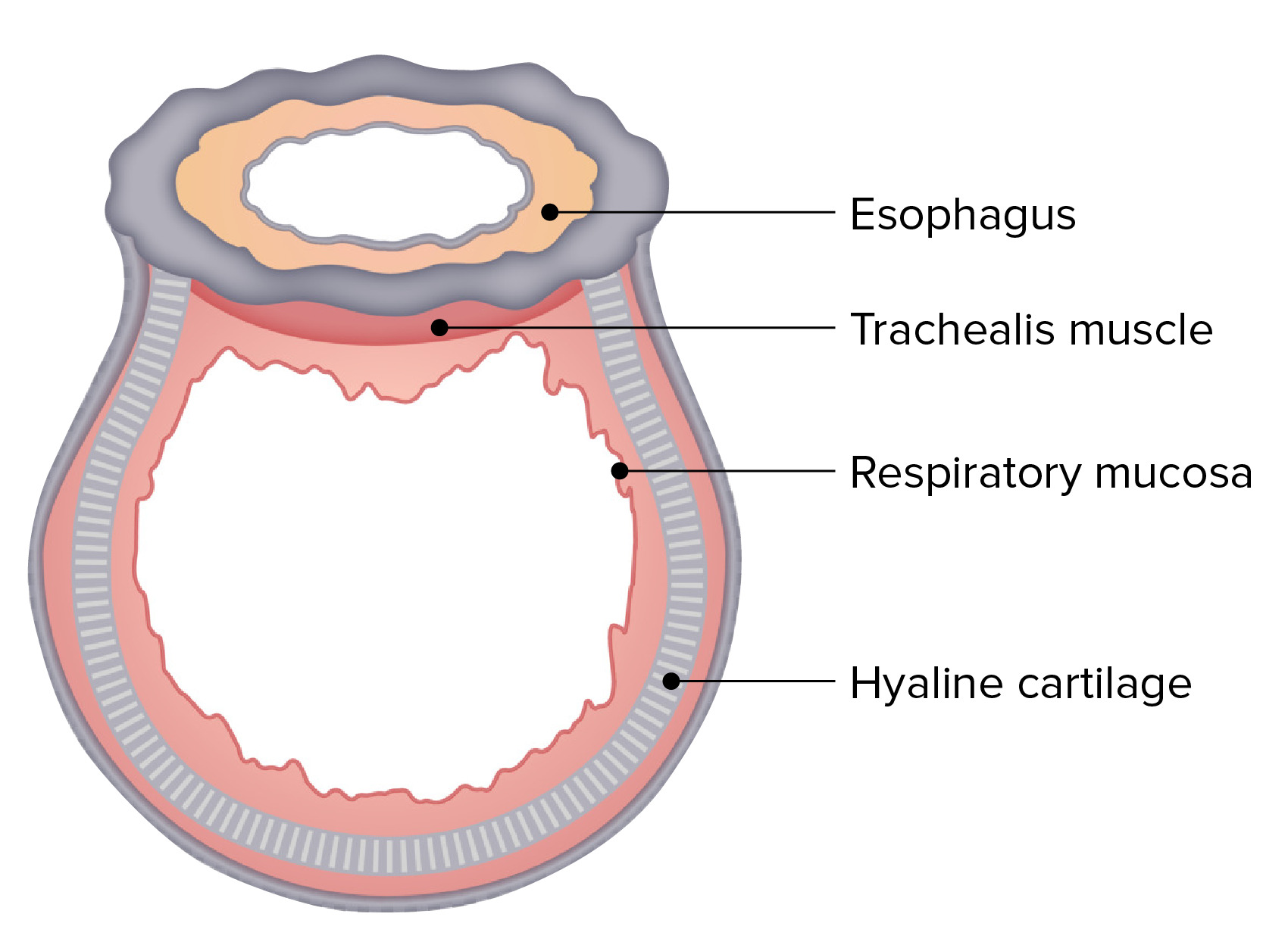
Which of the following statements about the pleura is FALSE?
- The pleura is lined by stratified columnar epithelium.
- The visceral and parietal pleura are fused around the hilum.
- The pleura is innervated by the intercostal and phrenic nerves.
- The pleura invaginates into two fissures in the right lung and into one in the left lung.
Which of the following statements about the pulmonary vasculature is FALSE?
- The pulmonary arteries transport oxygenated blood.
- The pulmonary veins transport oxygenated blood.
- The pulmonary arteries follow a similar branching pattern as the bronchi.
- The bronchial arteries supply blood to the airways.
Where do the pulmonary veins drain?
- The left atrium
- The right atrium
- The left ventricle
- The bronchial veins
- The right ventricle
The bronchial arteries provide blood supply to all of the following structures except...?
- ...the alveoli.
- ...the visceral pleura.
- ...the intrapulmonary blood vessel walls.
- ...the lymphatics.
- ...the terminal bronchioles.
Author of lecture Pulmonary Vasculature – Lung Anatomy

Jeremy Brown, PhD, MRCP(UK), MBBS
Customer reviews
2,3 of 5 stars
| 5 Stars |
|
0 |
| 4 Stars |
|
0 |
| 3 Stars |
|
2 |
| 2 Stars |
|
0 |
| 1 Star |
|
1 |
Very staggered - hard to listen to. It almost sounded like my laptop was lagging (which it wasn't because it's a brand new state-of-the-art laptop). This lecture needs to be redone.
The lecturer explained the content well but needs to enunciate better! I could barely understand some of the words.
reading off the slide, unclear explanation., unclear speech. Uses complicated terminology to explain simple ideas
