Playlist
Show Playlist
Hide Playlist
Introduction – Lung Anatomy


-
Slides 01 Respiratory Medicine Basics Brown.pdf
-
Download Lecture Overview
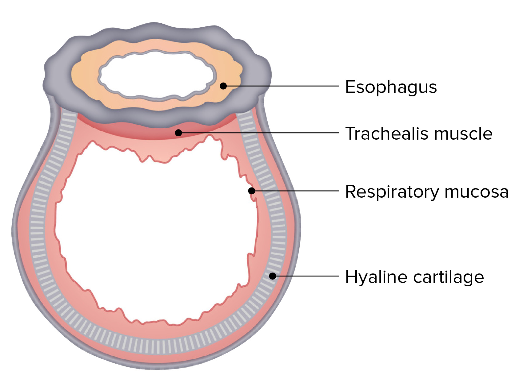
00:01 In the basic course on respiratory disease, I’ll discuss first the anatomy of the lung hoping to show you that the anatomy of the lung is dictated by its function. 00:10 Then I’ll describe the physiology of respiratory functions and then in the subsequent lectures I’ll discuss how the doctor approaches the patient’s to take a history, examine the patient and then thinks about and uses diagnostic tests to fully evaluate what their possible problem maybe that the patient is presenting with. 00:29 The anatomy of the lung includes the bones -- the vertebra, the ribs, the sternum; the lower airways -- the trachea, bronchial tree, bronchioles and alveoli; the blood supply to the lungs -- the pulmonary arteries, the veins, and bronchial circulation as well as lymphatic drainage; the muscles that move the lung -- the thoracic cage muscles, the intercostals, the diaphragm; the lining of the lung and the internal aspect of the thoracic cage -- the visceral and parietal pleura; and we will also touch on some of the nerves involved -- the phrenic and vagus nerve. 01:00 One aspect that we will discuss but is very important is that the respiratory tract actually starts in the nose so the upper airways of the respiratory tract as well that includes the nose, the mouth, the sinuses, the pharynx, the larynx and the vocal cords. 01:14 Right, so this diagram gives a general overview of the respiratory tract and you can see in the middle, the gray objects are the lungs -- the right lung on the right hand side, the left lung on the left hand side, and then nestling on either side of the heart and the pericardium. 01:29 And on the outside we have a layer of skin, muscle and then we have bones and you can see the pleura, which actually hangs down below both lungs on both sides. 01:41 And underneath the thoracic cavity we have the right and the left hemidiaphragm which separate the thoracic cavity from the abdomen and a vital for respiration. 01:52 Above the thoracic cavity we have the trachea which takes air in and out of the lungs and up into the upper airways in the neck. 02:00 This is a more schematic diagram showing the airways in entirety starting at the nose moving on to the pharynx which is at the back of the nose and then the larynx, the trachea, the bronchi and then the lungs at the end of the respiratory tract.
About the Lecture
The lecture Introduction – Lung Anatomy by Jeremy Brown, PhD, MRCP(UK), MBBS is from the course Introduction to the Respiratory System.
Included Quiz Questions
Which of the following is a part of the lower airway?
- Trachea
- Larynx
- Vocal cords
- Sinuses
- Pharynx
What divides the left lung into the superior and inferior lobes?
- The oblique fissure
- The horizontal fissure
- The transverse fissure
- The coronal fissure
- The oblong fissure
Author of lecture Introduction – Lung Anatomy

Jeremy Brown, PhD, MRCP(UK), MBBS
Customer reviews
5,0 of 5 stars
| 5 Stars |
|
1 |
| 4 Stars |
|
0 |
| 3 Stars |
|
0 |
| 2 Stars |
|
0 |
| 1 Star |
|
0 |
It was simply constructed well with diagrams and easily understood. He did not overcomplicate it.
