Playlist
Show Playlist
Hide Playlist
Esophagus, Stomach Dilation and Rotation, Duodenal Development

-
Slides 07-42 Esophagus, stomach dilation and rotation, duodenal development.pdf
-
Download Lecture Overview
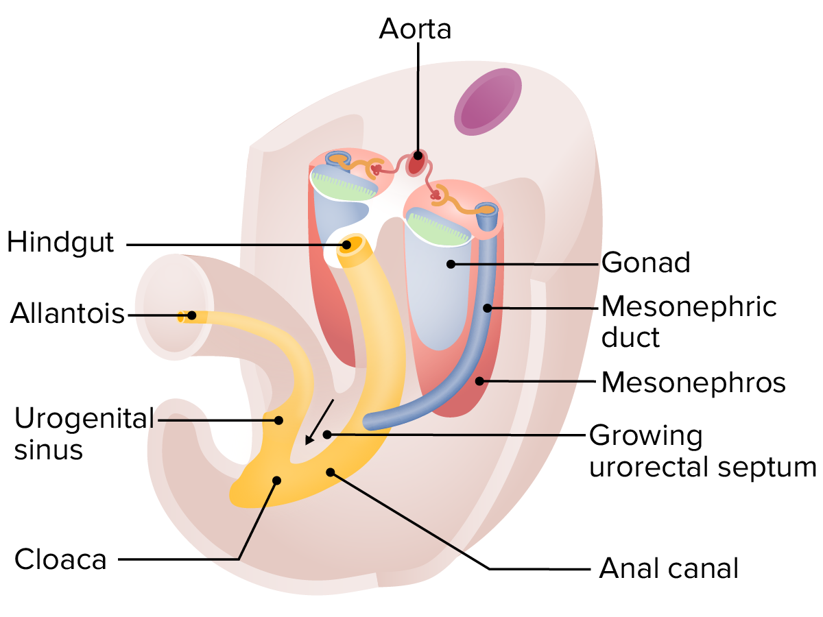
00:01 We’ll begin our exploration of how the foregut develops by looking at how the foregut and its associated glands separate from other organ systems. 00:09 Initially, the foregut is simply the area of the gut tube leading from the mouth a little further down to where the respiratory diverticulum buds off and later, the hepatic or liver bud is going to bud off of it as well. 00:22 Initially, it’s going to be a simple tube but will develop into the various foregut organs including the distal esophagus, the stomach, the liver, the gallbladder, the spleen, the proximal duodenum, and the large portion of the pancreas. 00:39 It receives its blood from the celiac trunk or celiac artery coming off of the aorta. 00:45 In the process of foregut development, we have to break it into multiple steps because there’s quite a lot going on. 00:52 In this talk, we’re gonna look at how the esophagus separates from the airway, the larynx and trachea, and then, follow how the stomach goes from a simple tube to ballooning out and rotating to create its mature shape. 01:05 Subsequent talks will look at how the liver, gallbladder, pancreas, and spleen develop from outgrowths of the gut tube from the foregut and then, how the foregut organs migrate as the liver, stomach, and other organs take their final position inside the abdominal cavity. 01:22 Now, this lecture is going to reiterate some of the points that were made when we discussed the respiratory system’s development and in particular, the development of the large airways. 01:33 Early on, we have a respiratory diverticulum growing off of the foregut and it is going to extend down to create the larynx, the trachea and the bronchi. 01:43 But to fully separate from the developing esophagus, we have a couple of tracheoesophageal ridges growing in from either side to meet on the midline to create a full tracheoesophageal septum and that’s what finally separates the airway anteriorly from the food carrying esophagus posteriorly. 02:06 Amongst things that can go wrong in the process of separating the airway from the esophagus are going to be tracheoesophageal fistulas or tracheoesophageal anomalies. 02:15 The most common version of this is gonna result in an esophageal atresia proximally and connection of the esophagus to the airway a bit more distally. 02:25 What’s going to happen in this case is that an infant who’s affected will attempt to feed but will then immediately regurgitate formula or milk because it literally has nowhere to go. 02:35 This could be diagnosed by trying to pass an oral gastric tube down the mouth and since it cannot get to the stomach, it will loop back and create a very distinctive looped shape on chest x-ray. 02:47 Other versions of tracheoesophageal fistulas can connect the esophagus to the airway and cause aspiration and may also be indicative of problems with the formation of the tracheoesophageal ridges. 03:00 Now, amongst other problems that can occur in this very proximal part of the foregut are gonna be congenital hiatal hernias. 03:07 This is simply the esophagus being too short. 03:10 So since the esophagus doesn’t reach the abdominal cavity, the very proximal portion of the stomach is inside the thoracic cavity and interestingly, many people have these and they are completely asymptomatic. 03:22 It’s not an uncommon finding amongst other things when you’re looking at chest x-rays to note that someone has a congenital hiatal hernia that they may be completely unaware of. 03:32 You wanna distinguish this from a sliding hiatal hernia where the esophagus is long enough to reach the abdomen but increased abdominal pressure pushes the very proximal stomach up into the thoracic area. 03:46 So it’s as though it’s just sliding through that opening of the esophagus into the abdominal cavity through the diaphragm. 03:54 It is important to distinguish congenital hiatal hernias and sliding hiatal hernias from a para-esophageal hernia. 04:00 In this case, the esophagus is travelling into the abdomen normally and does not slide in and out but rather, there’s a weak spot nearby that allows a portion of the stomach, likely, the fundus to herniate through alongside the esophagus. 04:15 This can occur congenitally and if so, can compress the lungs causing lung hypoplasia and not allow them to fill completely or develop normally. 04:23 We’ll now move on to how the stomach develops from a simple tube, into its interesting C-shaped and ballooned state. 04:32 The stomach begins by rotating in such a way that it’s left side moves anteriorly and its right side moves posteriorly. 04:40 So if I’m the stomach, something like this happens. 04:43 As that occurs, the stomach is going to twist around its longitudinal axis and will then start expanding posteriorly into the left. 04:52 Now, note here that the stomach is not hanging out in isolation. 04:58 It has a left vagus nerve and a right vagus nerve travelling down the esophagus to reach it and as it rotates, it’s going to pull that left vagus nerve anteriorly and the right vagus nerve posteriorly and that’s gonna create the left and right vagus nerve’s contribution to the anterior and posterior gastric nerves respectively. 05:21 Continued development of the stomach occurs when the side that’s facing to the left and slightly posteriorly balloons outwards and it balloons outward to a much more marked degree than the right side. 05:33 So instead of expanding uniformally, it balloons out to one side and stays pinched on the other and that’s what causes the stomach to adopt a relatively C-shaped appearance as it develops bringing its distal end no longer down but facing to the right. 05:51 Rotation of the midgut and migration of the liver are also involved in taking the stomach to its normal position on the left side of the abdomen but growth of its greater curvature on the left and failure of growth of its lesser curvature facing to the right and somewhat superiorly are gonna be what allows it to take on this ballooned but slightly unusual shape that we’re used to seeing. 06:15 The stomach’s pyloric region empties to the duodenum which will then have a C-shaped appearance and will be discussed further when we get to the midgut. 06:25 The pyloric region has a very thick smooth muscle sphincter within it and the pyloric sphincter is what normally allows food to stay in the stomach until it’s very gradually released as that sphincter relaxes. 06:40 Hypertrophy of the pyloric sphincter makes stomach emptying very difficult and we can see here on ultrasound, an extended pyloric sphincter with a very narrow lumen pointing down and a little bit to the left, and here we can see a barium swallow study with the fundus in the stomach very well represented and the stomach full of barium but a very, very thin trickle of barium through that stenotic pyloric canal. 07:09 This is typically something that develops between weeks two and six and is often described as a hard olive-shaped mass in the upper middle portion of the abdomen. 07:18 So it can be preliminarily diagnosed with palpation but you’d likely follow it up with an ultrasound or a barium swallow study to be definitive. 07:27 Moving into the duodenum, we mentioned in a prior talk that the duodenum is one place where we have a watershed between the superior mesenteric artery and the celiac artery. 07:39 Celiac artery for the foregut and superior mesenteric artery for the midgut. 07:43 Because there’s a failure of blood to supply this area cleanly, there can occasionally be some problems leading to atresia. 07:50 Another thing that makes the duodenum prone to trouble is that during normal development, its internal lining proliferates so tremendously that it actually blocks the actual lumen of the duodenum and it becomes impassible. 08:04 Normally, apoptosis is going to occur, the cells will die off and little vacuoles will form, enlarge, and eventually, fuse to recanalize the duodenum and if development proceeds normally, we have a complete continuous pathway from the stomach, through the duodenum, to the rest of the small intestine. 08:24 Sometimes these vacuoles do not form properly and they only partially recanalize the duodenum. 08:30 Other times, we can have narrowing of it, particularly, if there’s an ischemic event associated with the blood supply. 08:36 This makes travel of food through the duodenum difficult. 08:40 Even more extreme, we can have complete failure of recanalization because the vacuoles, spaces that form don’t connect or it’s so stenotic, it becomes atretic. 08:52 There’s actual atresia of the duodenum. 08:55 This is typically diagnosed through what’s known as the double bubble sign and it’s a funny name but a very good clinical finding to diagnose duodenal atresia since food and gas cannot pass through the midpoint of the duodenum, we tend to get gas bubbles trapped in the fundus of the stomach and the initial portion, the first portion of the duodenum and this double bubble is there because the gas and food cannot pass any further. 09:24 So double bubble sign can happen in any infant with duodenal atresia but is more common in those with Down syndrome. 09:32 Thank you very much for your attention, and we’ll follow-up on the rest of foregut development shortly.
About the Lecture
The lecture Esophagus, Stomach Dilation and Rotation, Duodenal Development by Peter Ward, PhD is from the course Development of the Abdominopelvic Region. It contains the following chapters:
- Development of the Foregut and Stomach
- Development of the Stomach
Included Quiz Questions
Which of the following is the most common tracheoesophageal anomaly?
- Proximal esophageal atresia with distal tracheoesophageal fistula
- Tracheoesophageal fistula
- Esophageal atresia alone
- Tracheal atresia alone
- Distal esophageal atresia with proximal tracheoesophageal fistula
Which of the following best describes a congenital hiatal hernia?
- When the developing esophagus does not lengthen sufficiently and pulls the cardiac region of the stomach into the thoracic cavity
- When the gastroesophageal junction moves freely through the diaphragm into the thorax as intra-abdominal pressure changes
- When the fundus of the stomach displaces through the diaphragm alongside the esophagus
- When the esophagus ends in a blind pouch, leading to the stomach entering the thoracic region
- When the hiatus muscle is not sufficiently developed, leading to free movement of the fundus into and out of the thoracic region
Which of the following statements regarding the development of the stomach from the gut tube is correct?
- As the stomach rotates, its left side becomes anterior and the right side moves to face posteriorly.
- The region of the foregut that becomes the stomach is initially distinct from the rest of the gut tube.
- As the stomach rotates, its left side becomes posterior and the right side moves to face anteriorly.
- During the development of the stomach from the foregut, there is little to no rotation.
- During rotation of the gut tube, the lower end of the stomach twists upward to become the gastroesophageal junction.
A newborn patient with poor stomach emptying, frequent, non-bilious vomiting and an 'olive-shaped' mass in the upper middle abdomen most likely has which of the following conditions?
- Narrowing at the opening of the pyloric sphincter
- Duodenal atresia
- Tracheoesophageal fistula
- Congenital hernia
- Stenosis of the small bowel
Which of the following is not associated with duodenal atresia?
- 'Olive-shaped' mass in the upper, middle abdomen
- 'Double-bubble sign'
- Trisomy 21
- Aspiration
- Duodenal epithelial proliferation resulting in blockage of the lumen
Author of lecture Esophagus, Stomach Dilation and Rotation, Duodenal Development

Peter Ward, PhD
Customer reviews
5,0 of 5 stars
| 5 Stars |
|
5 |
| 4 Stars |
|
0 |
| 3 Stars |
|
0 |
| 2 Stars |
|
0 |
| 1 Star |
|
0 |
