Playlist
Show Playlist
Hide Playlist
Bones and Muscles – Lung Anatomy


-
Slides 01 Respiratory Medicine Basics Brown.pdf
-
Download Lecture Overview
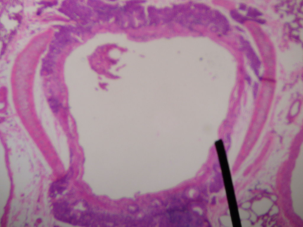
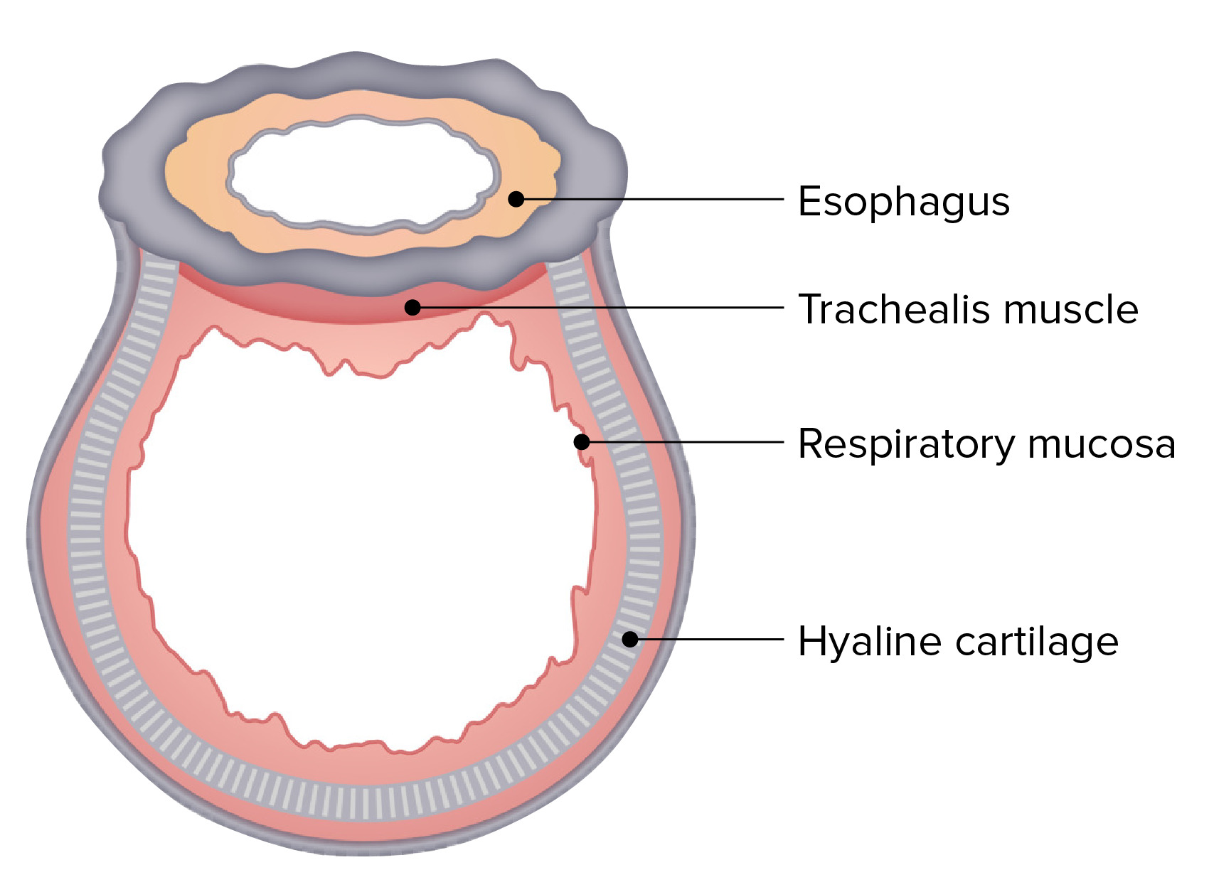
00:00 The lungs are kept within a articulated skeletal cage. And this is important, because it protects the lungs from damage, but also, it's important for function. Because it's expansion of the skeletal cage that allows the lungs to expand and take air in from the atmosphere. And this requires a fixed point. And there's a fixed point posteriorly, which is the thoracic vertebrae, and there are 12 thoracic vertebrae. Each is separated by cartilaginous intervertebral discs, and the vertebrae are the site of the articulation of the ribs, which curve around from the vertebrae to meet with a fixed point anteriorly, which is the sternum. And this is a flat bone, which you can all feel in the center of the anterior chest. And this comprises of three parts: the manubrium, which is the top part which joins to the clavicles, which are the bones that allow the shoulders and the sternum to be joined together; the sternal body, which is in the middle; and there's a little process that sticks out the bottom called the xiphoid process, and that's important because the diaphragm sticks onto that. The clinical relevance of this bony structure is that if you have several spinal defects—a curvature of the spine, or scoliosis—that will affect the mechanics of breathing, because that will affect the mechanics of expansion of the skeletal structures during respiration. And that can actually lead to respiratory failure if severe scoliosis is present. So the ribs are the bones that join the vertebrae and the sternum. And there are 12 pairs of ribs arising from the thoracic vertebrae, one from each. They're made of bone posteriorly, but anteriorly, they merge into cartilage, and it's these cartilages that form together to form the costochrondral margin, which you can feel between the abdomen and the thoracic cavity on both sides, and also merge to form with the sternum anteriorly (that's ribs 1 to 7). The clinical relevance of this is that the ribs articulate with the vertebrae, but those specific joints can be affected by a disease called ankylosing spondylitis. And if that disease is unchecked, then those articulations become rigid, and the ribs will not move during respiration. And again, that can lead to ventilatory defects, although it's a relatively rare cause. The 11th and 12th ribs are actually freestanding and are not directly involved in forming the thoracic cavity. An important thing about the ribs is that they… with each rib, there's an accompanying blood vessel—a vein and an artery—and also a nerve. The nerve supplies the skin overlying the rib. The vein and artery supplies the tissues around that rib. They all run together in a groove on the inferior surface of the rib, just inside. But it's important to know about that, because any procedure which involves putting needles or drains through the gaps between the ribs—an intercostal drain, for example—could penetrate the artery and cause bleeding, which very occasionally can be fatal. So it's important to know where those vessels run, and it's just underneath each rib. So this is an overview of the skeletal cavity or the thoracic cavity. You can see the sternum at the front of its body in the middle, the manubrium at the top, and the xiphoid process at the bottom. And the vertebrae run behind, and then the ribs run in between the two. To move the chest during respiration requires muscles. And there are three sets of muscles involved. There are the muscles of the chest wall: These are the serratus anterior and posterior, the trapezius of the neck, the pectoralis major and minor, rectus abdominis (the abdominal muscles down here). They are all involved in respiration when you're having forced respiration, such as when you're exercising, or if you have such severe lung disease that you need to maximize your ventilatory capacity. 03:55 The second set of muscles that are important are the intercostal muscles, and these are muscles that run between each rib. And they're divided into three categories, three types: there's external, which is on the outside; the internal is in between; and then the subcostal, which is the most inferior (they are closest to the parietal pleurae). And finally, there is the diaphragm. And so the intercostals are used for inspiration, and the diaphragm is used for inspiration as well. When you have forced expiration, i.e. when you're exercising, you'll also recruit these muscles for that process as well. 04:32 So to talk about the intercostals in a little bit more detail: These cross between each rib and are the main skeletal muscles of respiration. So the external ones, which are the outer layer—they run obliquely downwards anteriorly. That means that when they contract, they lift the ribs up and out and expand the chest. And they are one of the main drivers of inspiration. 04:52 The internals run obliquely downwards posteriorly, and they pull the ribcage back down again and therefore are used during forced expiration but are not necessarily used… are not necessary for normal expiration. The subcostal muscles run vertically, and they're in between… and they're the bottom layer, and they are actually underneath the intercostal vein, artery, and nerve. The clinical relevance of the intercostals is that if you have a problem affecting the skeletal muscles, such as motor neuron disease, then that can also lead to a lung hypoventilation (underventilation of the lung) and potentially respiratory failure. 05:29 And in fact, that is the mode of death for many patients with end-stage muscle disease, such as motor neuron disease or muscular dystrophies. This is a diagram, cross-section of the chest wall at the level of the diaphragm, and you can see here the intercostals, which are at the top part of this diagram with the external intercostal on the outside, the middle layer being the internal, and the underneath layer being the subcostal just next to the lung. 05:57 The diaphragm has that shape where it curves up between the lung and the liver on that side. On the left-hand side, it would be between the lung and the spleen and the stomach. And then there's an extra layers of skin and subcutaneous tissue and extrathoracic muscle outside of that. 06:15 The diaphragm itself is a smooth dome of skeletal muscle. 06:18 It’s placed under each lung, between the thoracic cavity and the abdomen. It has a central tendinous area, and it is essentially the main muscle of respiration. When it contracts, it makes the muscle smaller, and that pulls a domed diaphragm flatter, and that will expand the lungs downwards. The diaphragm arises from insertions on the xiphoid process anteriorly, the vertebrae back posteriorly, and the bottom six ribs around each side. In general, the upper limit of the diaphragm is on the 5th rib during… at rest and is slightly higher on the right-hand side than the left due to the presence of the liver beneath it on the right-hand side. Diaphragmatic movements and contractions are controlled by the phrenic nerve, which arises from the cervical 3, 4, and 5 nerve roots. 07:06 That's important, because without that phrenic nerve, the diaphragm will not move. If you have a bilateral phrenic nerve palsy—for example, if somebody has a cervical lesion above C3, 4, and 5 at C2—then they will die from respiratory failure unless there… some form of mechanical ventilation is used, because neither diaphragm is moving. There are three openings in the diaphragm, and these are important because they are sites of herniation. 07:29 So, for example, a hiatus hernia often comes up through the opening through the esophagus and the vagus posteriorly behind the heart and is visible on a chest x-ray. And the hiatus hernia represents stomach tissue… the stomach moving through the diaphragmatic opening into the thoracic cavity. The other openings are for the aorta and the inferior vena cava. 07:51 This is a diagram of the diaphragm. You can see the dome of the right and the left hemidiaphragm. 07:56 The sort of slightly gray area in the middle is the tendinous process. And you can see how the diaphragm arises posteriorly from the vertebrae of these insertions onto each vertebral body from the costochondral margin in the ribs around the side and the xiphoid process anteriorly. And this diagram clearly shows that sort of… that the dome shape of the diaphragm is, when it contracts, it will shorten, and that dome will flatten. 08:19 And that's expands the lungs.
About the Lecture
The lecture Bones and Muscles – Lung Anatomy by Jeremy Brown, PhD, MRCP(UK), MBBS is from the course Introduction to the Respiratory System.
Included Quiz Questions
Which of the following statements about the chest wall is FALSE?
- The sternum is a single bone.
- There are twelve pairs of ribs.
- The anterior cartilaginous ends of ribs 1 to 7 fuse with the sternum.
- The intercostal artery and nerve run along the inferior aspect of each rib.
How many thoracic vertebrae are present in the body?
- 12
- 9
- 10
- 14
- 15
The xiphoid process is the anterior attachment point for which of the following structures?
- The diaphragm
- The pericardium
- The intercostal muscles
- The visceral pleura
What is scoliosis?
- Sideways curvature of the spine
- Reduced conduction through the phrenic nerve
- Exaggerated medullary respiration signaling
- Pathological straightening of the spine
- Decreased curvature of the thoracic spine
Author of lecture Bones and Muscles – Lung Anatomy

Jeremy Brown, PhD, MRCP(UK), MBBS
Customer reviews
4,5 of 5 stars
| 5 Stars |
|
1 |
| 4 Stars |
|
1 |
| 3 Stars |
|
0 |
| 2 Stars |
|
0 |
| 1 Star |
|
0 |
it is not so clear for me it was ok and can be improved
1 customer review without text
1 user review without text
